The Malykhin Lab

The Malykhin Lab

Neuroimaging · Computational Neuroanatomy · Human Brain

Gallery

The lab in pictures

The people behind the work, alongside a selection of figures from the lab’s published research.

Lab life

Life in the lab

The group today, and moments from the lab’s earlier years on campus and on the road.

2025–2026

The current team
The lab team seated in front of the bear sculpture
By the bearFall 2026 · University of Alberta
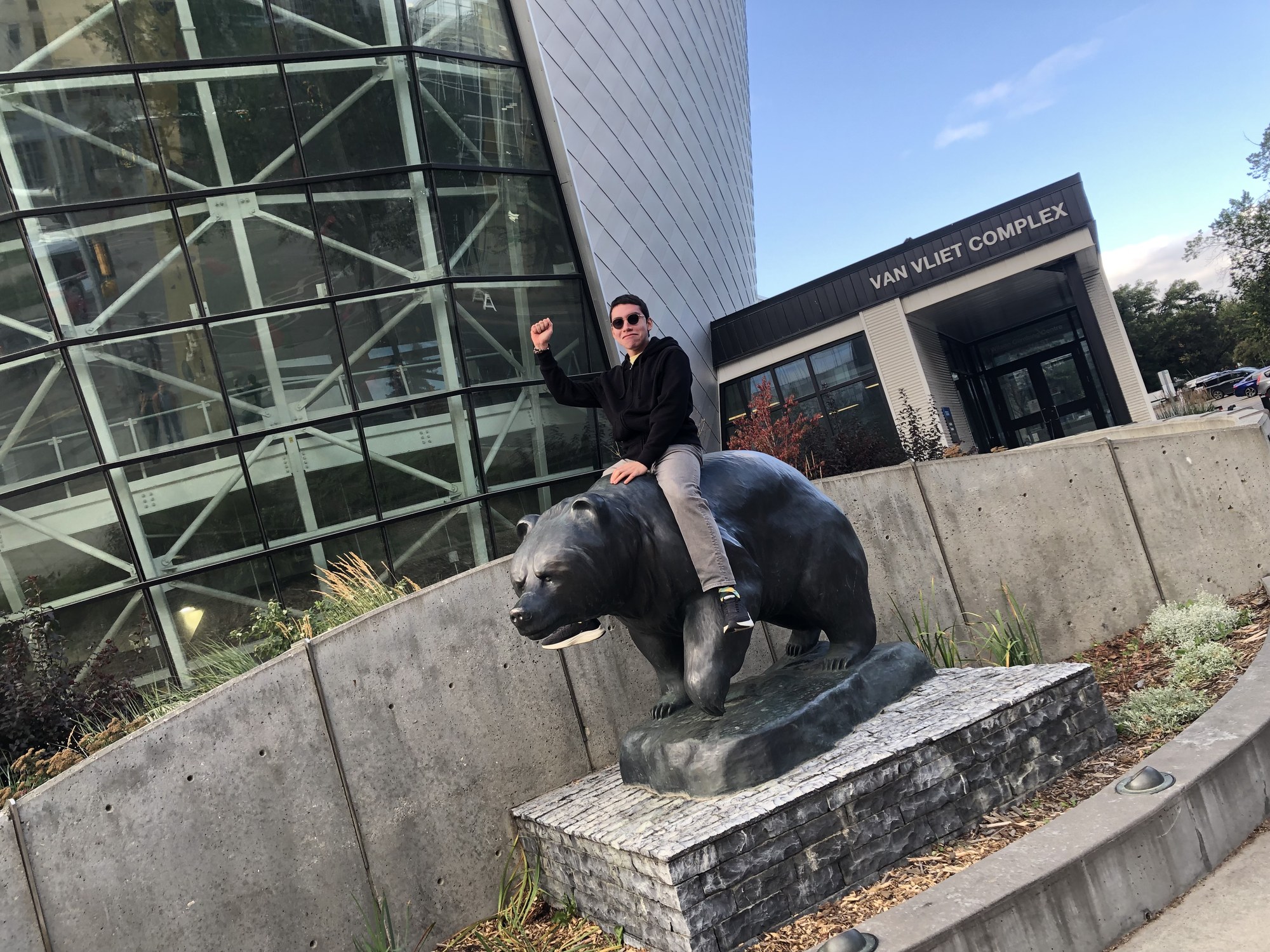
Ethan Hernandez sitting on the bear sculpture outside the Van Vliet Complex
Ethan rides the bearFall 2026 · Van Vliet Complex
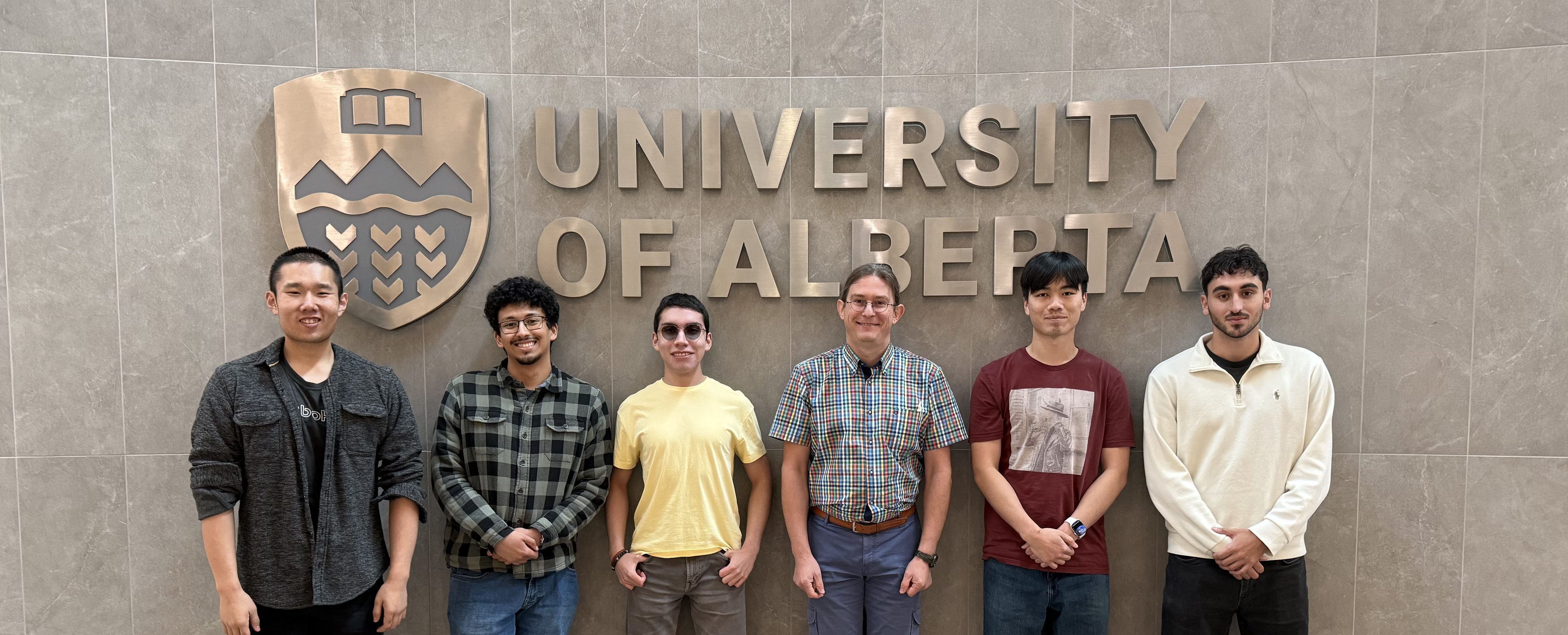
The lab team in front of the University of Alberta crest
At the crestFall 2026 · University of Alberta
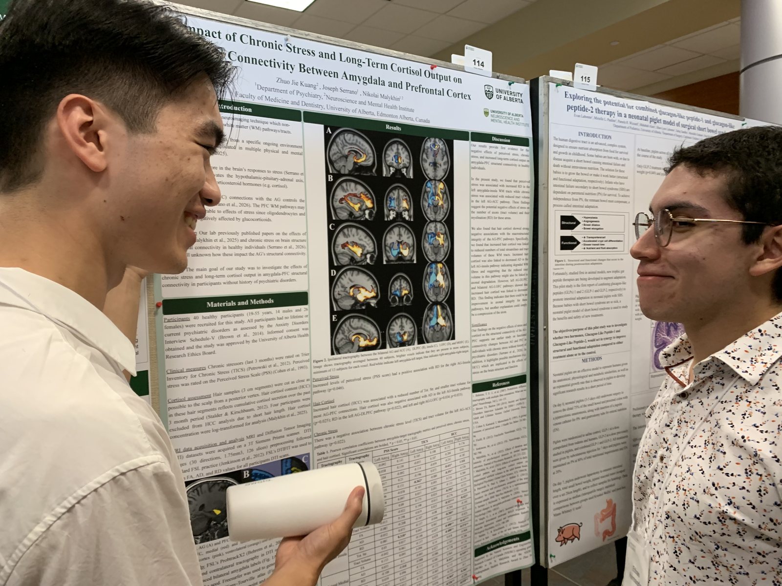
Jacky and Ethan at the poster session
FoMD Research DayAugust 2026
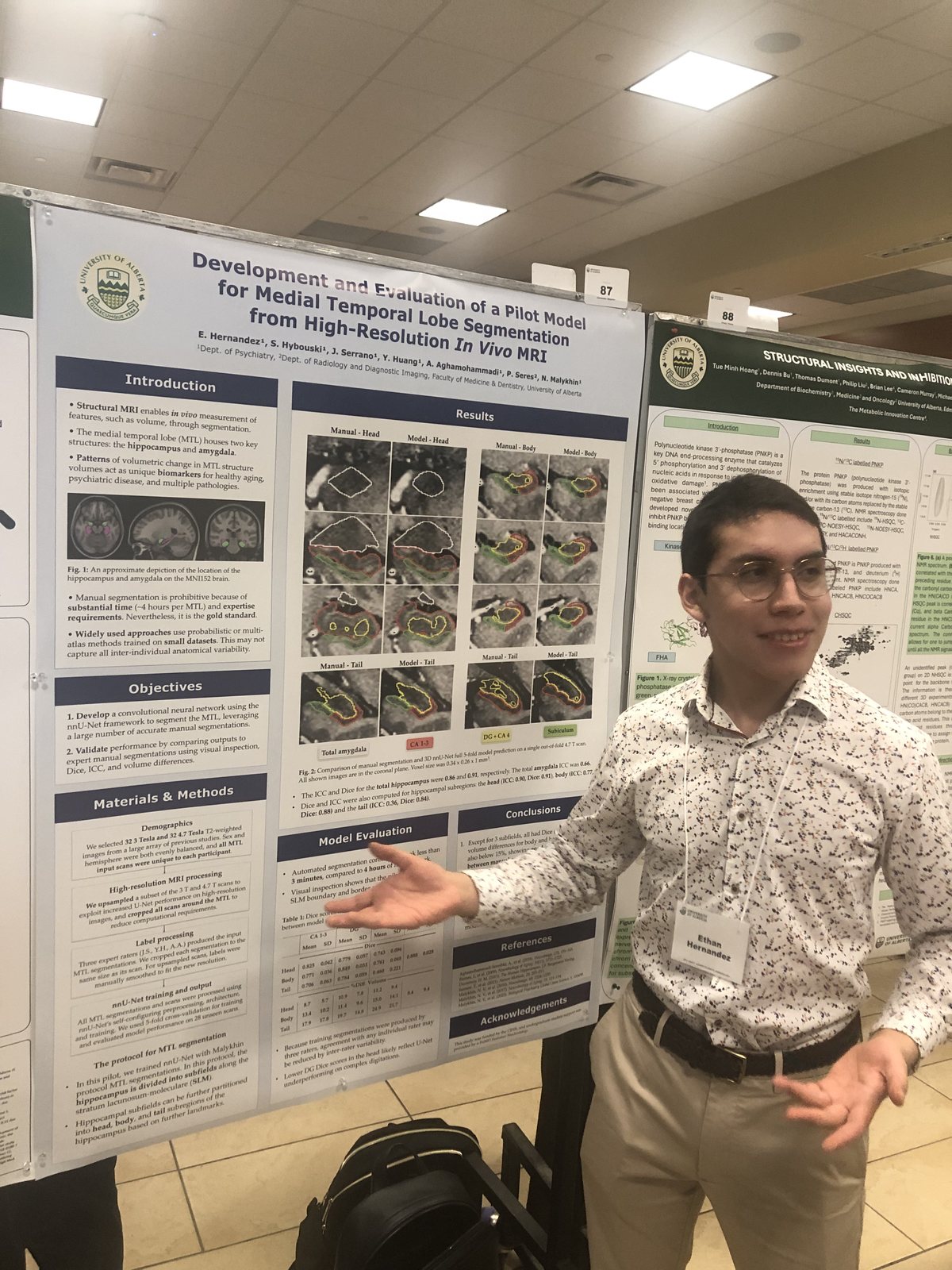
Ethan Hernandez presenting his poster
Ethan presentsFoMD Research Day
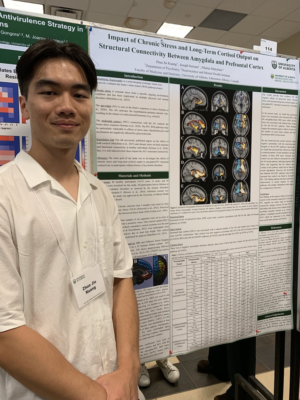
Jacky Kuang beside his poster
Jacky presentsFoMD Research Day
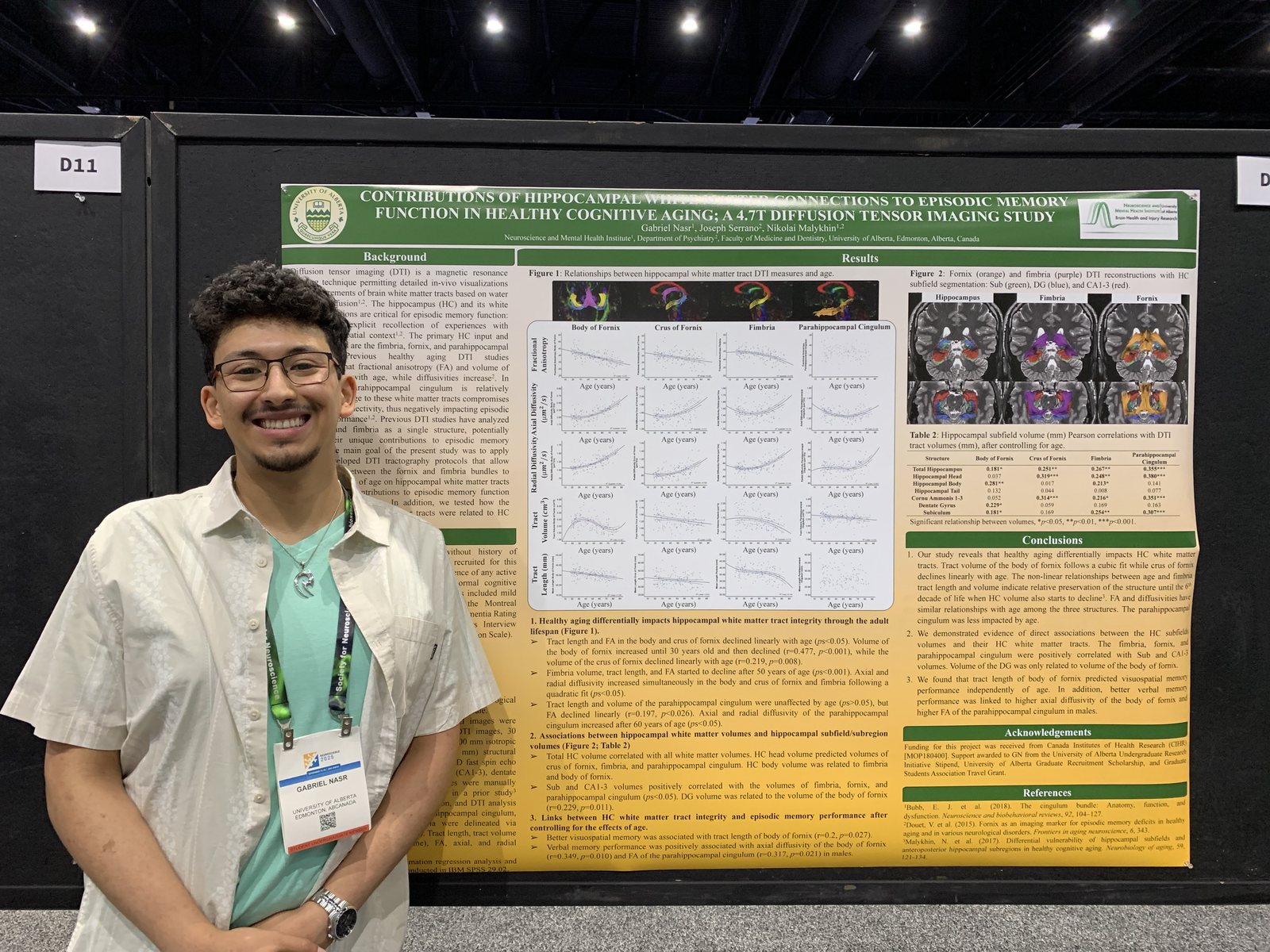
Gabriel Nasr beside his poster at Neuroscience 2025 in San Diego
Gabriel at Neuroscience 2025Society for Neuroscience · San Diego · November 2025Contributions of hippocampal white matter connections to episodic memory function in healthy cognitive aging: a 4.7 T diffusion tensor imaging study

Earlier years

On campus and on the road
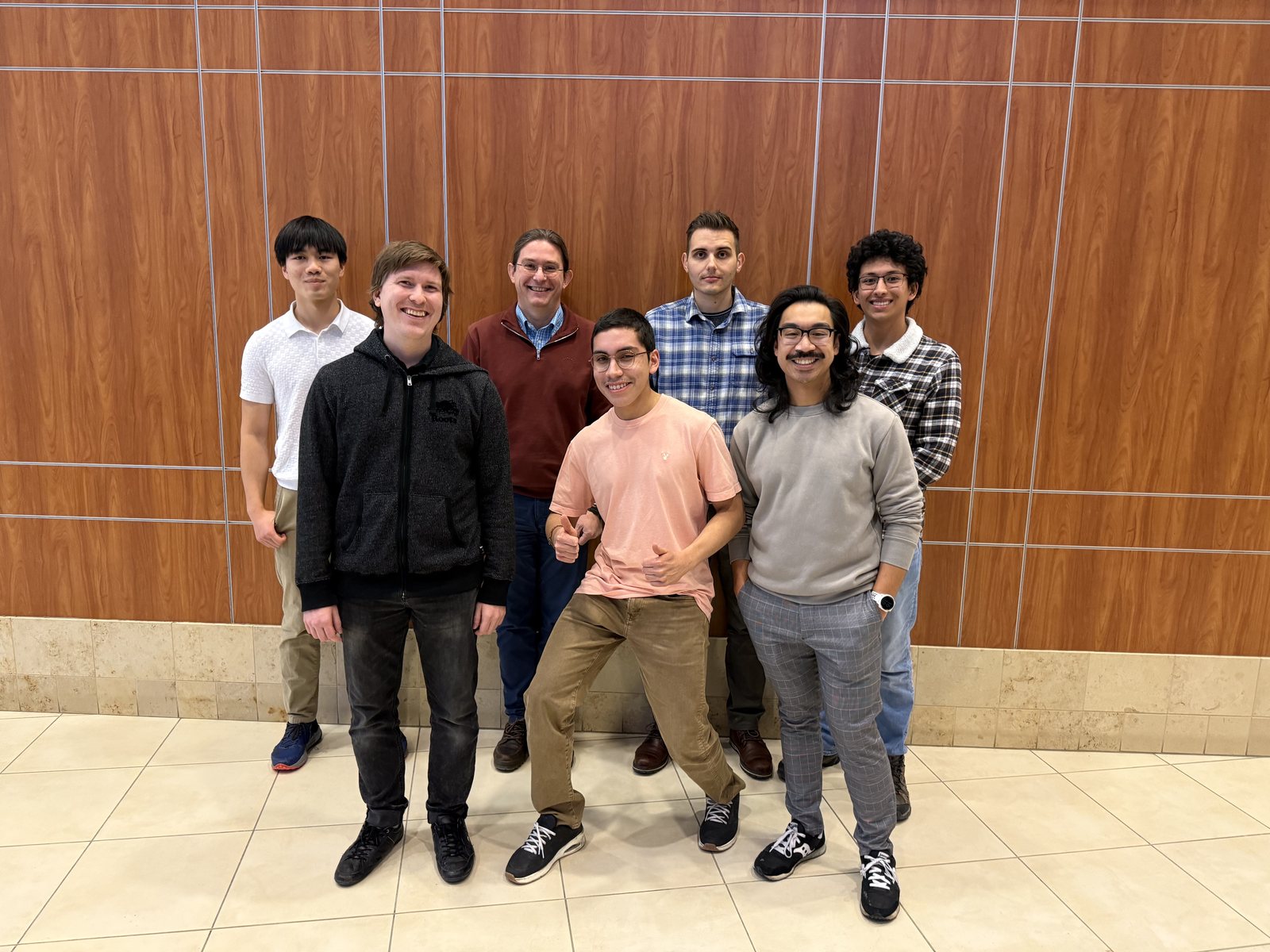
The Malykhin Lab team in fall 2024
The lab in fall 2024University of Alberta
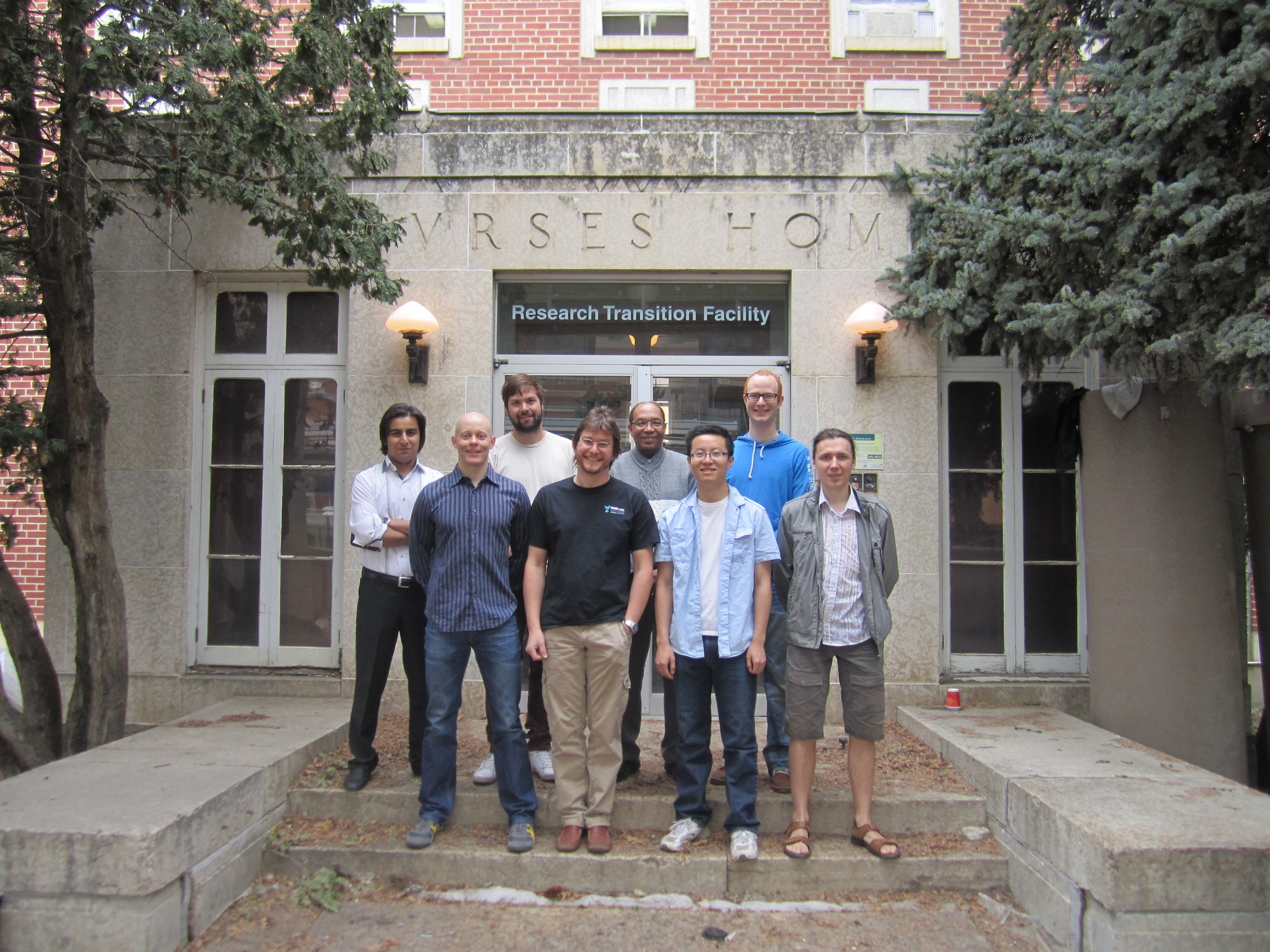
Lab members on the steps of the Research Transition Facility
At the Research Transition FacilityUniversity of Alberta
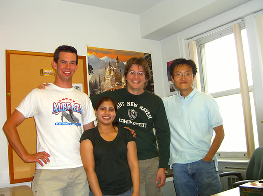
Lab members together in the office
In the labUniversity of Alberta
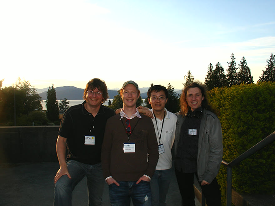
Lab members outdoors at a conference by the coast
On the roadConference travel
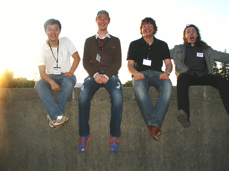
Lab members together at sunset
Good companyAfter hours
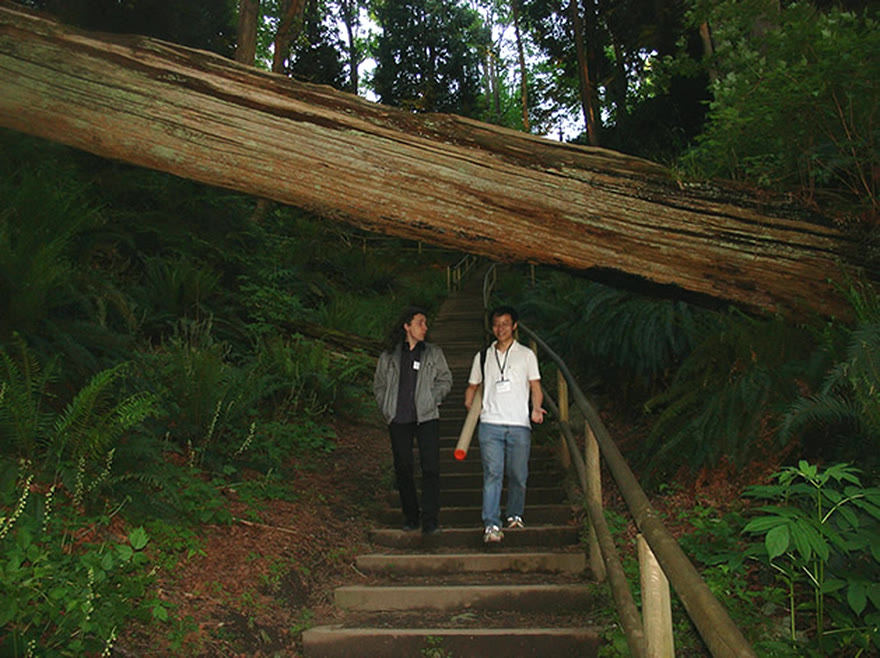
Two lab members walking a forest trail
Into the woodsWest coast
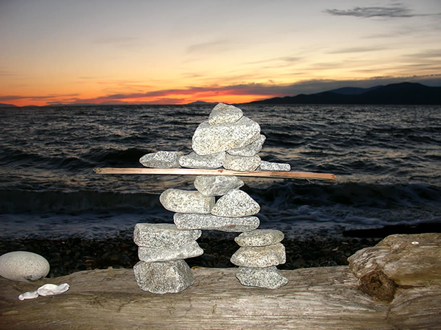
An inukshuk on the beach at sunset
Sunset by the waterWest coast
In the lab

Work from the lab

Reconstructions, segmentations, and analyses produced from the lab’s own MRI data. Select any image to see it in more detail.
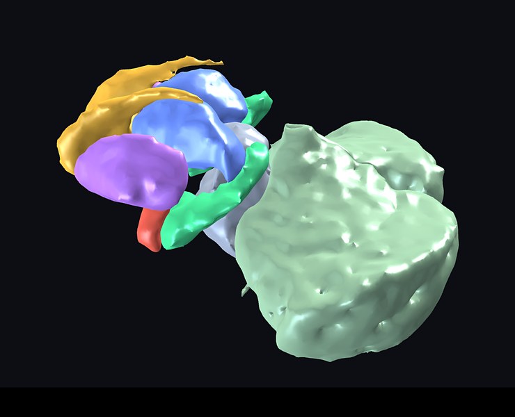
Whole-brain atlas showing the subcortical structures, brainstem and cerebellum
Whole-brain atlasSubcortical structures, brainstem and cerebellum from the interactive 3D atlas
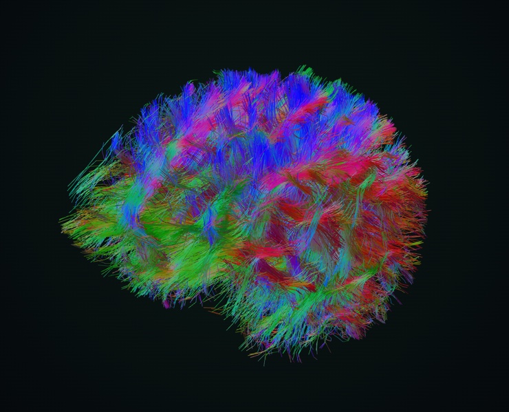
Whole-brain tractography
Whole-brain tractographyFibre pathways from diffusion MRI, coloured by orientation
Hippocampus and amygdala in 3D
Hippocampus and amygdala in 3DManual segmentation on 4.7 T fast spin-echo MRI