Neuroscience: A Journey Through the Brain
 |
Neuroscience: A Journey Through the Brain |
| Main Page Organization Development Neuron Systems About the site (Glossary) References |
The Major Systems: Sensory Systems - Visual System
|
Sensory System sub categories: Chemical Visual Auditory Somatic |
For a human to 'see' stimuli in their external environment, several processes must occur:
Focusing the image on the retina (back to top)
The human eye collects light rays emitted or reflected off objects in the environments, and focuses them onto the retina to form images. Several structures in the eye contribute to this function:
| Structure | Function |
| Cornea | Refracts light rays, focusing them on a specific point on the retina |
| Lens | Assists the cornea in refracting light to focus an image by accommodating (becoming rounder as ciliary muscles contract) |
| Pupil | Continually adjusts for ambient light levels via the pupillary light reflex |
Visual Transduction (back to top)
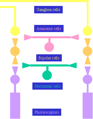
Once the image on the retina has been formed, the 'neuroscience' part of vision can begin: visual transduction. The cells in the retina are organized inside-out: the first cells to receive stimulation are the innermost photoreceptors, and the cells that exit the eye to carry information to the brain are the outermost cells. The following diagram depicts the cellular arrangement in the retina:
 |
The organization of the retina is termed laminar (meaning 'layered'). The layers are named as follows:
The photoreceptors are of two types: rods and cones. Aside from structural differences, these two types of receptors also possess functional differences. Rods are adapted to vision in low light, while cones are adapted to high acuity, color vision in high light conditions. Photoreceptors indicate light when they are hyperpolarized by light, and the transduced signal exits the eye via the ganglion cell axons, forming the optic nerve. |
Integration of visual information in the brain (back to top)
There are two major channels of information flow from the retina to the striate (or visual) cortex. The M-path runs from the M-type ganglion cells to layers 1 and 2 of the lateral geniculate nucleus (LGN), located in the thalamus. From there, projections are made to layer IVCa of the striate cortex. This pathway is specialized for detecting movement, orientation and direction.
The P-path runs from the P-type ganglion cells to layers 3 to 6 of the LGN, then on to layer IVCb of the striate cortex. This path is specialized for detecting color and object form.
Created and Maintained by: Melissa
Davies
Last Updated: April 09, 2002 09:02 PM