Neuroscience: A Journey Through the Brain
 |
Neuroscience: A Journey Through the Brain |
| Main Page Organization Development Neuron Systems About the site (Glossary) References |
Neural Induction and Neurulation
|
Development Home Neural Induction Diversification Migration Synapse Formation |
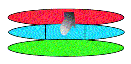
In the human embryo, there are 3 layers of tissue from which body organs are derived: endoderm, mesoderm, and ectoderm. Endodermal tissue forms the gut, lungs and liver; mesodermal tissue forms muscles, bones, and vasculature; and ectodermal tissue forms the nervous system and the epidermis.
Jump to section: Mesodermal Induction | Neurulation | Brain Development
|
Mesodermal Induction (back to top) A series of 3 experiments provided evidence for the role of mesodermal tissue in inducing the differentiation of ectodermal tissue to form the nervous system. Removing the mesoderm prevented ectoderm differentiation. The central part of the mesoderm causes CNS differentiation, as when it was removed, the CNS did not develop. Furthermore, there is a rostral-caudal gradient in the mesoderm. Transplanting two sections of rostral mesoderm resulted in the formation of two forebrains from the ectoderm. The mesoderm releases factors that activate promoter or repressor sites on genes in the ectoderm. Typically, a gradient is seen for these factors along the rostral-caudal gradient in the mesoderm. |
 |
|
Neurulation (back to top) The animation shows how the primitive layers develop into the spinal cord (part of the CNS). As the ectoderm (yellow) begins to acquire neural properties, it forms the neural plate (red). Ectoderm that does not follow the neural differentiation path gives rise to the epidermis of the skin. The neural plate begins to fold, creating the neural groove, which develops into a tubular structure called the neural tube. This process is called neurulation. The caudal region of the neural tube gives rise to the spinal cord (shown in animation), while the rostral region gives rise to the brain. The center of the tube is called the central canal, and is filled with CSF. Future muscle cells arise from somites, which in turn arise from the mesoderm (blue). Endoderm is shown in green. |
 |
|
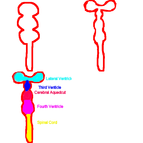
Brain Development (back to top) The neural tube gives rise to the ventricular system, which is surrounded by neural tissue (shown in red). Initially, the neural tube is divided into 3 sections: the forebrain, midbrain and hindbrain. As the nervous system develops, the forebrain is further divided into the telencephelon and diencephelon; the midbrain is also referred to as the mesencephelon; and the hindbrain is further divided into the metencephelon and myelencephelon. The telencephelon surrounds the lateral ventricles (light blue). The diencephelon surrounds the third ventricle (dark blue). The mesencephelon surrounds the cerebral aqueduct (pink). The metencephelon and myelencephelon surround the fourth ventricle (green). The spinal cord is filled with CSF in the central canal (yellow). Failure of the caudal portion of the neural tube to close results in Spina Bifida. Failure of the rostral portion of the neural tube to close results in Anencephaly, which is usually lethal. Obstruction of the cerebral aqueduct result sin build-up of CSF in the lateral and third ventricles and causes Hydrocephalus. |
 |
Created and Maintained by: Melissa
Davies
Last Updated: April 09, 2002 08:56 PM