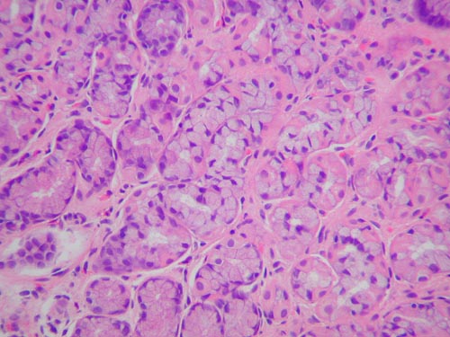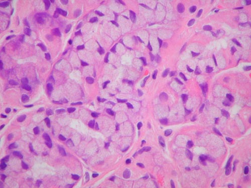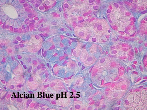
I was obviously concerned about the possibility of signet ring carcinoma in situ in this patient. In view of the normal endoscopy, negative family history, lack of significant atypia and the presence of similar alcian blue positive cells in other patients I would be happy with a rescope and additional biopsies of any suspicious areas (see followup below). Assuming there is no evidence of malignancy I will assume the changes are reactive until I can prove otherwise. It would be interesting to measure his serum gastrin level.
Comparison with a known signet ring carcinoma with adjacent signet ring carcinoma in situ has been provided by Gerard Abadjian from last year. I am not surprised that signet ring carcinomas are felt to originate from mucous neck cells after having viewed the changes seen in our patient.


Authors
Konda Y. Kamimura H. Yokota H. Hayashi N.
Sugano K. Takeuchi T.
Title
Gastrin stimulates the growth of gastric pit with less-differentiated
features.
Source
American Journal of Physiology. 277(4 Pt 1):G773-84, 1999
Oct.
Abstract
Gastrin stimulates the growth of gastric mucosa by increasing
mostly its
glandular region but is not known to induce the growth of a
pit region
where its major constituent cells, gastric surface mucous (GSM)
cells,
turn over rapidly. To investigate the effect of gastrin on GSM
cells, we
generated hypergastrinemic mice by expressing a human gastrin
transgene.
We obtained a hypergastrinemic mouse line whose average serum
gastrin
level is 671 +/- 252 pg/ml (normal level <150 pg/ml). Gastrin-positive
cells were found in the fundic mucosa. The gastric mucosa exhibited
hypertrophic growth, which was characterized by an elongated
pit with an
active proliferative zone, but the glandular region containing
parietal
cells was normal or reduced in size. The GSM cells contained
fewer mucous
granules than those of control littermates and lost reactivity
to the GSM
cell-specific cholera toxin beta-subunit lectin. GSM cells along
the
foveolar region and many mucous neck cells became Alcian blue
positive,
suggesting the appearance of sialomucin in these cells. We suggest
that
gastrin stimulates the growth of the proliferative zone of gastric
glands,
which results in the elongation of the pit region whose GSM
cells exhibit
less-differentiated features.
Followup
All those who responded felt the changes were metaplastic.
Vann Schaffner told us that the late Roger Haggitt reviewed a similar case
and came to the same conclusion. Our patient went for repeat gastroscopy
with multiple biopsies which showed the same changes in body
mucosa. There was no evidence of invasion or atypia.
For what its worth I have since found a textbook reference in the Fenoglio
CD-ROM which states that the mucous neck cells in the
normal stomach "sometimes produce small amounts of sialomucins and sulfomucins"
but is no more specific than that and uses the following references.
Suganuma T, Katsuyama T, Tsukahara M, et al: Comparative histochemical
study
of alimentary tracts with special reference to the mucous neck cells
of the
stomach. Am J Anat 161:219, 1981.
Filipe MI: Mucins in the gastro-intestinal epithelium. A review. Invest
Cell
Pathol 2:195, 1979.