1. What are the properties of the magnocellular and parvocellular layers of the lateral geniculate nucleus (LGN)?
2. Describe the structure and function of early visual pathways.
3. How is the visual cortex organized?
4. What are the properties of the simple, complex, and hypercomplex/end-stopped cells in the visual cortex?
5. What are the functions of some extrastriate areas?
6. What are the two different extrastriate pathways? Describe the evidence that supports this distinction.
7. What do visual agnosias and blindsight reveal about visual processing?
- optic nerves from from retinal ganglion cells (RGCs) meet at optic ______ (crossover point)
e.g., object in the left visual field:
- image passes through lens and projects on the right part of the retina in both eyes
- at optic chiasm, optic nerves from right side of both eyes meet
- optic ______ (optic nerve after the optic chiasm) project to the LGN in the thalamus
- then to the right half of the visual cortex in the occipital lobe
- layers of lateral __________ nucleus (LGN) of the thalamus (sensory processing centre):
parvocellular |
magnocellular |
|
RGC input: |
P-cells |
M-cells |
cell body size: |
small |
large |
number: |
many |
few |
conduction speed: |
slow |
rapid |
response type: |
sustained |
transient |
receptive field size: |
small |
large |
percepts: |
high _______ detail |
______ sensitive |
colour: |
colour |
black & white |
Two pathways:
1. _______________ visual pathway
- P-cells, some M-cells (P and M pathways) → LGN
- LGN has 6 layers; each has a ___________ map: adjacent neurons correspond to spatially related points on the retina
(LGN also receives substantial feedback connections from the cortex)
- then, optic radiations project to cortex in _________ lobe:
• primary visual cortex (V1, “_______ cortex”)
• and secondary visual cortex (V2)
2. _____________ visual pathway
- remaining M-cells → superior colliculi of the ______: part of brain stem; guide visual attention
- then projects to thalamus: ________ and lateral posterior nuclei, then to V2 and beyond
- controls eye movements/fixations; detection/orientation to visual stimuli, motion and location
- has 6 layers (magno → 4Cα, parvo → 4Cβ)
- layers 2 and 3 contain:
- _____: sensitive to wavelength, but not orientation (mostly receive input from parvo)
- __________: areas between blobs; sensitive to orientation, but not wavelength (receive input from parvo only)
David Hubel & Torsten Wiesel (1962, 1968):
- shared 1981 Nobel Prize for research on information processing by cortical cells in the visual system
- found cells in V1 that responded well to certain stimuli:
• ______ cell (layer 4) responds best to:
▸ a bar, line, or edge of light
▸ in a particular location on the retina
▸ having a specific orientation
• _______ cell (layers 2/3 and 6) responds best to:
▸ a bar, line or edge of light
▸ in a particular location on the retina
▸ having a specific orientation
▸ and moving in a certain direction
• ____________/end-stopped cell (beyond V1) responds best to:
▸ a bar, corner, or angle having a certain length and/or width
▸ in a particular location on the retina
▸ having a specific orientation
▸ moving in a certain direction
- when inserting electrode perpendicular to surface of cortex, cells responded to stimuli having the same property
• ________ column: cells respond to stimuli from same retinal location
• ______ dominance column: cells respond to stimuli presented to one eye only
• ___________ column: cells respond to line stimuli having the same orientation; adjacent orientation columns differ in orientation selectivity by 10°
• ___________: region containing a single location column, which contains left and right ocular dominance columns, which contain the set of orientation columns from 0° to 180°
Occipital Lobe
• V3: cells sensitive to ______ edges of a certain orientation
- believed to handle perception of _____ and local motion
- projects to temporal and parietal lobes
• V4: cells respond to perceived ______ of a surface (not wavelength)
Verrey (1888):
▸ 61-year-old female stroke patient with cerebral _____________: unable to perceive colour in the right half of her visual field
▸ could perceive but not identify different colours
e.g., purple and orange look different, but colours could not be identified
- projects to temporal lobe
Temporal Lobe
• ________ temporal (IT) cortex: involved in identifying stimuli
Gross, Rocha-Miranda, & Bender (1972):
▸ single-cell recording in monkeys
▸ waved _______ to unresponsive cell → fired!
▸ cells responded best to silhouette of a monkey’s ___
- _______ cells: respond best to simple stimuli, such as dots, squares, ellipses
- _________ cells: respond best to more complex shapes, or shapes combined with colour or texture
- face detector cells discovered via _____________: inability to recognize faces due to IT damage (fusiform gyrus)
e.g., The Man Who Mistook His Wife For a Hat (Sacks, 1985)
• ______ temporal (MT) cortex (a.k.a. V5): sensitive to overall motion (and direction) of object--but not colour
Zihl, von Cramon, & Mai (1983):
▸ 43-year-old female: small lesion due to vascular disorder
▸ cerebral ___________ (a.k.a. motion agnosia): unable to see objects in motion
- projects to parietal lobe
Two somewhat distinct neural pathways (Ungerleider & Mishkin, 1982):
• _______ (or temporal) pathway
- parvo → V1 → V2 → V4 → IT
- concerned with object recognition and ______________
- a.k.a. “____” system
• ______ (or parietal) pathway
- magno → V1→ V2 → V3 → V4, & MT (V5) → parietal lobe
- involved in ________ objects, motion, spatial relationships, depth
- a.k.a. “_____” system
Evidence:
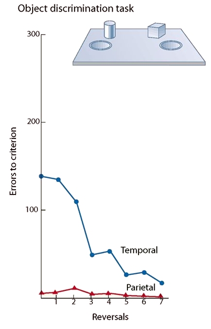
Pohl (1973): lesion studies
- monkeys given delayed response tasks:
• ______ discrimination: shown object (e.g., brick), then presented choice task (brick and cylinder): if target (brick) was moved, it would uncover a hidden well that held food reward
• ________ discrimination: one object presented; food hidden in well closest to object
- monkeys trained to criterion on one of these tasks, then task was reversed
- after learning, either temporal or parietal lobe lesioned
- effects of lesion on object task:

• temporal-lobe lesion monkeys show much impairment
• parietal-lobe lesion monkeys show minimal impairment
• suggests “____” pathway affected
- effects of lesion on landmark task:
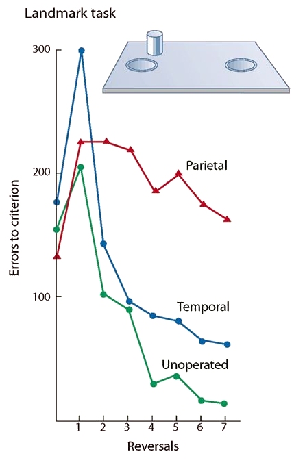
• unoperated monkeys show no impairment
• temporal-lobe lesion monkeys show minimal impairment
• parietal-lobe lesion monkeys show much impairment
• suggests “_____” pathway affected
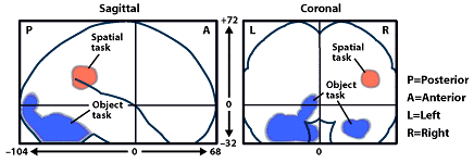
Kohler et al. (1995): human neuroimaging
- observers presented two frames in succession, with a brief delay in between
- objects were moved or replaced with other objects
- ______ task: same objects?
- _______ task: same locations?
- data indicate evidence for what-where distinction:
Visual agnosia: failure or deficit in perceiving or recognizing visual objects (Freud, 1891); from Greek, meaning “without knowledge”
e.g., _____________: disruption of face perception; inability to recognize/identify friends and family; inability to read facial expressions/emotions
• may not be able to perceive a face
• or may have intact perception of facial features, but inability to identify face
Categories of visual agnosias (Lissauer, 1890):
• ____________: failure to form a holistic percept; deficit in perception of whole objects
- inability to extract global structure, despite intact low-level sensory processing (acuity, colour, and brightness discrimination intact)
- cannot recognize, discriminate, or copy complex visual forms, like ______
- but they can grasp objects they cannot identify
- a.k.a. visual form agnosia, or visual space agnosia
- neuropathy: caused by diffuse lesions to posterior occipital cortex (e.g., due to CO or mercury poisoning)
- likely a failure of “binding” of features together, due to damage at early stage of visual processing
• ___________: deficit in associating percept with meaning ("recognition without meaning")
- cannot draw from memory
- able to copy pictures, but cannot ________ them
- can use other senses (e.g., touch, smell)
e.g., visual object agnosia: “convoluted red form with a linear green attachment” = ____ (Sacks, 1970)
- neuropathy is not consistent; different subtypes may involve different perceptual impairments
- whole object not identified, likely due to damage to later stage of visual processing that connects visual object to semantic knowledge
Specific visual agnosias (Farah, 2004):
• ________ specific agnosia: inability to identify living (or nonliving) objects, metals, fruit, vegetables, musical instruments, fabrics, or gemstones
• ___________ agnosia: able to recognize drawings of objects rotated in picture-plane, but impaired at recognizing picture’s orientation; drawings often copied perpendicular to original
• _______________: inability to perceive more than one aspect of a visual stimulus and integrate details into coherent whole
- dorsal: can only perceive one of a number of ___________ objects; unattended objects not perceived; often cannot localize objects; bilateral damage to occipitoparietal regions
- ventral: can perceive more than one object at a time, but cannot identify more than one; may be able to describe one aspect of a scene without understanding the _____; can localize objects; damage to left inferior temporo-occipital cortex
• pure ______: cannot read words; must do letter-by-letter reading; can write to dictation but cannot read back what has been written; can copy words, which leads to recognition
- may be the same as _______ simultanagnosia
• ___________ agnosia: impaired recognition of scenes and landmarks; get lost easily; damage to inferior medial occipito-temporal cortex (similar to prosopagnosia)
Double-Dissociation of Extrastriate Pathways: in one case study, one ability is functioning, but another ability is not; and vice-versa in another case study
________ syndrome (Rudolf Bálint, 1909):
- patient had bilateral damage to superior posterior parietal lobes (very rare)
- oculomotor ______: inability to reach for and grasp objects in field of view
- optic _______: inability to guide eye movements or change visual fixation
- simultanagnosia
- could recognize individual objects, but could not tell where they were located or reach for them
- intact “what” system, but damaged “_____” system?
Milner & Goodale (1996):
- patient D.F. (“Diane”) had CO poisoning, which led to occipitotemporal brain damage (ventral stream)
- visual form agnosia: unable to discriminate object identity, shape, orientation
- cannot recognize or draw an apple or a book by looking at them--but can draw them from memory or recognize by holding objects
- can perceive colours and textures
e.g., she can identify a banana from its yellow colour and texture of its surface)
- to her own ________, she can accurately reach and grasp objects
e.g., can reach out to appropriately grasp a banana
- tested on two similar, but different tasks:
• ___________ matching: “Hold the card at the same slant as the slot”; D.F. much worse than controls
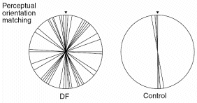
• visuomotor _______: “Put the card into the slot”; D.F. about the same as controls (intact dorsal stream)
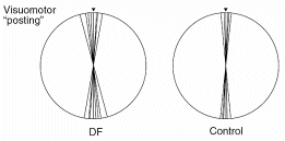
- damaged “what” system, but intact “where” system?
- maybe not “what” vs. “where”, but “what” vs. “___” (how to interact with objects in space)
Weiskrantz & colleagues (1974):
- patient D.B. had right _______ cortex removed to treat abnormal blood vessels
- stimuli could not be detected if they fell on the right half of either retina (vision was excellent for the other half-retinas)
- however, D.B. could accurately point to spots of light, even though he insisted that he did not actually ___ them
- could accurately guess whether a stick was vertical or horizontal, but had no idea whether he was right or wrong “because I couldn’t see anything; I couldn’t see a ____ thing”
- some patients can even correctly identify facial expressions 90% of the time
- associated with V1 damage
- due to intact pathway(s) to extrastriate areas bypassing V1:
• _____________ pathway
• possible LGN → V5 pathway
- implies some visual information is processed at an ___________ level; but geniculostriate pathway is responsible for conscious vision