J Pharm Pharmaceut Sci (www.ualberta.ca/~csps) 7(1):27-36, 2004
Systemic bone formation with weekly PTH administration in ovariectomized rats.
Sébastien A. Gittens1,2#, Gregory R. Wohl3#, Ronald F. Zernicke4, John R. Matyas5, Paul Morley6,Hasan Uludag1-3*
1 Faculty of Pharmacy & Pharmaceutical Sciences, 2 Department of Biomedical Engineering, Faculty of Medicine, 3 Department of Chemical & Materials Engineering, Faculty of Engineering, University of Alberta, Edmonton, Alberta, Canada, 4 Faculty of Kinesiology, 5 Department of Cell Biology & Anatomy, Faculty of Medicine, University of Calgary, Alberta, Canada, and 6 Institute for Biological Sciences, National Research Council of Canada, Ottawa, Ontario, CanadaReceived 17 December 2003, Revised 23 January 2004, Accepted 23 January 2004, Published 30 January 2004
PDF Version
Abstract
PURPOSE: Weekly subcutaneous administration of 0 (vehicle), 10 and 80 mg/kg doses of human parathyroid hormone (1-34) [PTH (1-34)] were compared based on their capacity to induce systemic formation of bone in 9 month-old ovariectomized (OVX) Sprague-Dawley rats. METHODS: Changes elicited at bone tissue after 4 weeks of treatment were assessed using dual x-ray absorptiometry, micro-computed tomography (mCT), and ashing. RESULTS: The 10 mg/kg dose led to a significant increase (p<0.025) in femoral bone mineral density (BMD) over vehicle- and 80 mg/kg-treated groups. Similarly, structural analysis of the femoral neck trabecular bone by mCT revealed increases in bone volume fraction and trabecular thickness over the pre-treatment baseline, and vehicle- and 80 mg/kg-treated groups. CONCLUSIONS: The data suggest that the weekly administration of 10 mg/kg of PTH (1-34) was sufficient to significantly promote the bone mineral density systemically. The weekly administration of 10 mg/kg over a 4-week treatment period is, to our knowledge, one of the lowest reported total dose of PTH (1-34) shown to induce a net anabolic effect on skeletal tissue in OVX rats.
Introduction
The continuous administration of the amino-terminal fragment (1-34) of parathyroid hormone [PTH (1-34)] has been shown to elicit a catabolic effect on skeletal tissue. Intermittent administration of PTH (1-34), however, has been shown to promote the deposition of new bone [1-3]. Although the biochemical pathways responsible for mediating these divergent responses have yet to be fully elucidated [2], the anabolic effects of PTH (1-34) has been observed in normal rats [4-7] as well as the rats rendered osteopenic by neurectomy [8], tail-suspension, [8-10], aging [11-17], streptozotocin-induced diabetes [18], and orchidectomy [19]. Since the ovariectomized (OVX) rat is one of two animal models mandated by the US Food and Drug Administration for the preclinical assessment of agents designed to treat osteoporosis [20], much of the literature concerning the effects of PTH (1-34) administration on systemic bone regeneration has been based on this particular model. As assessed through x-ray absorptiometry, histomorphometry, micro-computed tomography (mCT), and various other techniques, the preponderance of the evidence provided by studies using this particular animal model suggests that the daily, parenteral administration of PTH (1-34) resulted in an anabolic effect on the surfaces of both trabecular and cortical bone [21-24]. The predominant corollary associated with PTH (1-34) administration is not only the regeneration of mineralized tissue on the axial and appendicular skeleton, but an improvement in bone architecture in terms of increased connectivity, trabecular thickness, etc. It has been shown that the culmination of these effects is an enhancement in bone biomechanical performance [25, 26].
Despite these encouraging results, recent reports suggest that long-term administration of the synthetic PTH (1-34) was associated with some detrimental side effects [27, 28]. For example, Sato et al. reported that the daily administration of as little of 8 m g/kg PTH (1-34) for 1 year in OVX rats led to an 11% increase in brittleness (i.e. ultimate displacement) of diaphyseal cortical bone and a 48% reduction in diaphyseal marrow space [27]. In addition to these adverse skeletal effects, exogenous PTH therapy has been associated with hypercalcemia both in animal studies, as well as in clinical studies [29]. Besides the hypercalcemia, transient headaches, nausea and anthralgia were also reported with clinical PTH (1-34) administration [29].
It was thought, therefore, that a reduction in the dosing frequency could serve as a means of circumventing some of these adverse effects. Consequently, the aim of this was to investigate the anabolic response elicited by low dose PTH (1-34) administered weekly. The doses selected in this study, as well as the administration duration were considerably lower than the other studies reported in the literature. It was our desire to determine whether such a restricted dosing regimen would have had any systemic effects on skeletal tissues.
Materials and Methods
Animal Care
Twenty-seven, 2 month-old, commercially ovariectomized, female Sprague-Dawley rats were purchased from Charles River Laboratories (Quebec City, PQ). Rats were acclimated until 9 months of age under standard laboratory conditions (23°C, 12 h of light/day) prior to the beginning of the study. While maintained in pairs in sterilized cages, rats were provided standard commercial rat chow and tap water (ad libitum). All procedures involving the rats were approved by the Animal Welfare Committee at the University of Alberta (Edmonton, Alberta).
Experimental Design
Rats were weighed and assigned randomly into four groups. Rats in the initial baseline control group (n = 6) were killed at the beginning of the study just prior to the onset of the treatment protocols. For 4 weeks, the remaining 3 groups (n = 7 per group) received subcutaneous injections once per week of either vehicle (acidified saline), a low PTH (1-34) dose (10 mg/kg per injection), or a high PTH (1-34) dose (80 mg/kg per injection). Eight days after the final injection rats were killed via asphyxiation using a CO2 chamber and weighed. At autopsy, the uterus was excised and weighed to confirm systemic depletion of estrogen. Bone tissues including both femora, tibiae, and a portion of the lumbar spine (L1-L4), were harvested at time of euthanasia, fixed immediately in 70% ethanol, and stored at -20°C.
The delivery vehicle used consisted of saline (0.9 % NaCl; Baxter Corporation, Toronto, ON) acidified to 0.01 mM HCl. The synthetic hPTH (1-34) (lot #: NO5129A1, American Peptide Company, Sunnyvale, CA) was diluted in the delivery vehicle just prior to weekly injections.
Bone Analysis
Femur Length: Using a vernier caliper (reading error ± 0.01mm, Mituyomo, Japan), femoral bone length was assessed as the distance between the proximal and distal femoral growth plates - a measure of femoral diaphyseal length.
Mineral Ash Content: Mineral ash content was determined by ashing 1-cm long segments of tibia from the proximal tibial growth plate to the mid-diaphysis [30]. Samples were defatted in acetone and then dried for 24 hr at 100°C. The dry samples were weighed in air (dry bone mass), and then rehydrated in distilled water for 24 hr under vacuum and weighed underwater (wet bone mass). The samples were then ashed at 800°C for 24 hr and weighed (mineral ash mass). Archimedes principle was used to determine bone density (r, g/cm3 ) and volume (cm3 ). Ash content was calculated as a measure of mineral ash mass per dry bone mass (ash content, %), and mineral ash mass per bone volume (ash density, g/cm3 ).
Femoral Bone Mineral Density (BMD): Femoral BMD (g/cm2 ) was measured by dual-energy X-ray absorptiometry (DEXA; Piximus Mouse Densitometer, GE Medical Systems Canada, Ottawa, ON). BMD, calculated by dividing bone mineral content (g) by the projected bone area (cm2 ), was assessed for the total femur as well as for three equivalent femoral sub regions: the proximal third, central third, and distal third of each femur.
Trabecular Bone Architectural Properties: To elucidate the anabolic effects of weekly PTH (1-34) administration, structural parameters of the trabecular bone within the femoral neck were evaluated through mCT. Architectural changes in the trabecular bone of the rat femoral neck and L3 vertebrae were specifically analyzed (Skyscan 1072 X-ray Microtomograph, Skyscan, Aartselaar, Belgium). All bone samples were scanned at 100 kV/98 mA. The proximal femurs were scanned at an isometric resolution of 11.46 mm/pixel and 11.46 mm slice thickness, and the L3 vertebra were scanned at an isometric resolution of 14.89 mm/pixel with 14.89 slice thickness. Reconstruction of the original scan data was performed using a cone-beam algorithm [31] into two-dimensional 1024 X 1024, 8-bit, 256 grayscale bitmap image slices. For the femoral neck, identically sized cylindrical volumes of interest (VOI) were sampled from bitmap images stacks to include trabecular bone regions and exclude cortical bone within each femoral neck data set. Each VOI of femoral neck trabecular bone was centered within the femoral neck about the narrowest region, approximately 2 mm distal to the femoral head growth plate, and was oriented parallel to the axis of the femoral neck. Likewise, ellipsoid VOI were sampled from each L3 bitmap data set to include only trabecular bone within the vertebral centrum. The axis of the L3 cylindrical VOI was aligned along the cranial-caudal axis of the vertebral centrum. Digital segmentation of the bone from air/marrow tissues was performed by global thresholding. The global threshold for each sample data set was determined by selecting a local minimum in the frequency plot of bitmap pixel grey levels for the sample [32]. The VOI were then reconstructed in three dimensions (ANT, Skyscan, Aartselaar, Belgium), and measures of bone volume fraction (BV/TV), bone surface fraction (BS/BV), and trabecular thickness (Tb.Th.). Trabecular number was calculated separately (3-D Calculator v0.9, Skyscan, Belgium)
Cortical Bone Cross-section (XSA): Cortical bone cross-sectional areal properties were measured by analyzing single mCT bitmap cross-sectional image slices of the rat femoral necks. The BITMAP images were sampled in a plane perpendicular to the femoral neck axis at the narrowest portion of the femoral neck. Each slice was analyzed using custom software (MATLAB, ver.6.0.0.88, rel.12, The Mathworks, Inc., Natick, MA) for measures of femoral neck cross-sectional area (XSA), and moments of area about the horizontal (Ixx) and vertical axes (Iyy) for the whole bone and separately for the cortical bone and trabecular bone. XSA measures were also calculated for the area within the periosteal envelope (i.e., femoral neck XSA enveloped by the periosteal perimeter including all bone and myeloid spaces), and the endosteal envelope (i.e., the XSA enveloped by the endosteal perimeter including trabecular bone and marrow spaces).
Statistical analysis
Unless otherwise noted, all values are expressed as mean ± standard deviation. Rat body masses were compared between the onset and end of treatment within each animal using student's paired t-test (SPSS for Windows 11.0.1, SPSS Inc., Chicago, IL). For comparisons of all other variables among treatment groups, significant main effects were tested by multivariate analysis (generalized linear model). Where main effects were significant (p<0.05), treatment groups were compared using Tukey post-hoc comparison (p<0.05). To test whether or not either rat body mass or uterine mass had a confounding effect on bone properties, all data were also compared by multivariate analysis with rat body mass and uterine mass as covariates.
Results
Measures of femoral BMD and tibial ash content were not obtained for the baseline (pre-treatment) group. All other measures ( mCT based bone properties, rat body mass, and uterine mass) were compared among all treatment groups including the baseline group.
Body Mass, Uterine Mass, and Femoral Diaphyseal Length
Body mass did not differ among the treatment groups either at the onset of the treatment protocols (9 months, p=0.537), or at termination (4 week into treatment, p=0.193). There was no significant change in body mass over the 4 wk treatment interval. Ovariectomy was confirmed by reduced uterine masses of the rats at sacrifice (0.15 ± 0.08 g, n=27; historical data from our lab indicated uterine mass of ~0.9 g for the age-matched normal rats). There were no differences in uterine mass among the treatment groups. A comparison of the groups with rat body mass and uterine mass as covariates found that neither parameter had any significant interaction on ash content, bone mineral density, or bone architectural properties. The femoral diaphyseal lengths did not differ significantly (p=0.185) among the baseline group (34.4±1.0 mm), the vehicle group (34.5±1.2 mm), the low-dose PTH (1-34) group (35.7±0.7 mm), or the high-dose PTH (1-34) group (34.9±1.0 mm).
Tibial Ash Content
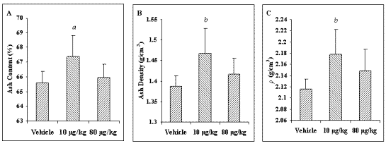
To assess changes elicited by weekly PTH (1-34) administration on the bone mineral content, segments of the proximal tibia were ashed. Weekly treatment of 10 mg/kg PTH (1-34) resulted in significant increases in both ash content (2.7%) and ash density (5.8%) compared to the vehicle-treated group (Figure 1).
Figure 1: The effects of weekly PTH (1-34) administration on bone mineral content in the tibia. Tibial ash content is presented as a function of either dry weight (A), or volume (B). Bone density of the segmented samples was also determined (C). In all cases, the weekly administration of 10 mg/kg PTH (1-34) led to a statistically significant increase over the vehicle-treated group. ap<0.025 vs. vehicle-treated group. bp<0.0125 vs. vehicle-treated group.
In addition, an increase of 2.9% in tibial bone density was observed in the 10 mg/kg PTH-treated group compared to the vehicle-treated group. Slight increases in the same parameters were observed in high-dose PTH group, but these values were not significantly different from the vehicle-treated controls.
Femoral Bone Mineral Density
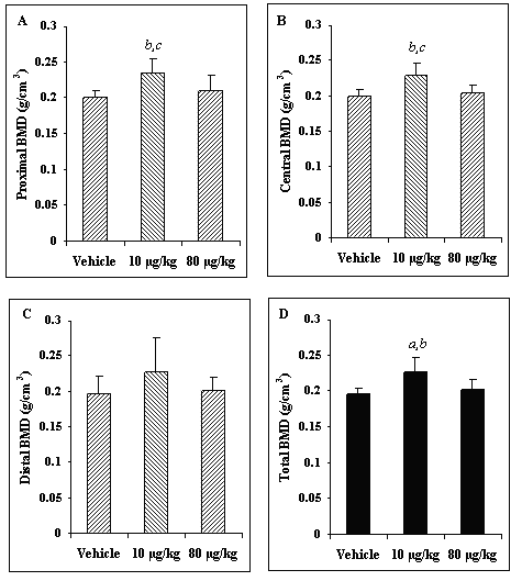
Weekly administration of low-dose PTH led to a significant (p<0.0125) increase of 15.6% in total femoral BMD over the vehicle-treated controls (Figure 2). Weekly low-dose PTH also led to a significant increase of 8.9% in BMD over the high-dose PTH group. Both the proximal and the mid-diaphyseal regions of the femur exhibited similar, statistically significant increases in the 10 mg/kg-treated group over the vehicle and high-dose PTH groups. Although a similar trend was observed in the distal region of the femur, these differences were not statistically significant (p=0.222).
Figure 2: The effects of weekly PTH (1-34) administration on bone mineral density within the proximal (A), central (B), and distal (C) regions as well as the total (D) femur. In all of the selected regions, except distal, a significant increase in the 10 mg/kg-treated group was observed over that of the vehicle- and 80 mg/kg-treated groups. ap<0.05 vs. 80 mg/kg-treated group. bp<0.025 vs. vehicle-treated group. cp<0.025 vs. 80 mg/kg -treated group.
Architecture of Trabecular Bone within the Femoral Neck
As summarized in Figure 3, treatment by vehicle alone had no appreciable effects on femoral neck trabecular bone architectural properties compared to baseline control.
Figure 3: The effect of weekly PTH (1-34) administration on structural parameters BV/TV (A), BS/TV (B) and Tb.Th. (C), of the trabecular bone within the femoral neck. Listed in the table is a summary of all of the architectural parameters generated for this region. ap<0.05 vs. 80 mg/kg-treated group. bp<0.0125 vs. vehicle and 80 mg/kg-treated groups and. cp<0.0125 vs. all other groups.
Weekly low-dose PTH (1-34) treatment did result in significant increases in BV/TV and Tb.Th. (p<0.0125) in the femoral neck over all other groups including baseline. BS/BV, on the other hand, was significantly reduced in the femoral necks of 10 mg/kg-treated animals compared to all other groups. Despite a significant reduction in Tb.N. in the 10 mg/kg-treated group over baseline group (p<0.05), no other statistically significant differences were found for either Tb.N. or Tb.Sp among the 3 study groups. Altogether, these data suggest that a net anabolic effect had been elicited by the weekly administration of 10 mg/kg PTH (1-34) on the trabecular bone within the femoral neck.
Femoral Neck Bone Cross Sectional Area (XSA)
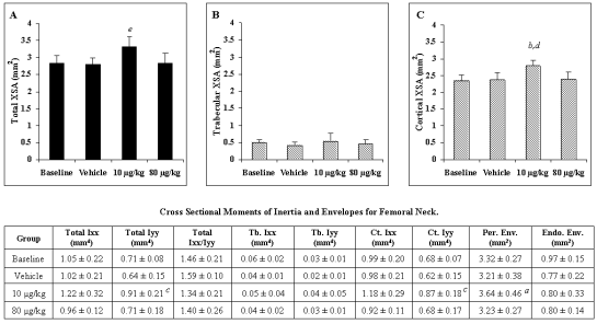
Total XSA of the femoral neck was significantly larger in the 10 mg/kg-treated group compared to all other groups (Figure 4).
Figure 4: The effect of weekly PTH (1-34) administration on total (A), trabecular (B) and cortical (C) XSA within the femoral neck. Summarized in the table are the second moments of area about the horizontal and vertical axes (Iyy and Ixx, respectively) for total, trabecular (Tb.) and cortical (Ct.) XSA as well as the areas of the periosteal and endosteal envelopes (Endo.Env. and Per.Env., respectively). ap<0.05 vs. vehicle-treated group. bp<0.05 vs. 80 mg/kg-treated group. cp<0.025 vs. vehicle-treated group. dp<0.0125 vs. baseline and vehicle-treated groups. e p<0.0125 vs. all other groups.
When XSA area was evaluated separately as cortical and trabecular regions, the statistically significant increase was found for the cortical, but not the trabecular bone. Specifically, the 10 mg/kg cortical XSA was significantly greater than the cortical XSA in all other test groups (p<0.0125). Further analysis revealed that PTH treatment with 10 mg/kg significantly increased the XSA area within the periosteal envelope compared to treatment by vehicle alone (data not shown). However, XSA within the endosteal envelope tended to decrease in all treated groups compared to baseline, there were no significant differences among the groups (data not shown). A significant increase of 13% (p<0.05) in the area of the periosteal envelope was found between the 10 mg/kg PTH (1-34)-treated group over the vehicle-treated group. Despite a 17% reduction in the 10 mg/kg PTH (1-34)- compared to vehicle-treated group, changes in the area of the endosteal surfaces were not significant among any of the groups. The culmination of these data suggested that the weekly administration of 10 mg/kg PTH (1-34) influenced the cortical bone within the femoral neck in an anabolic manner.
The second moments of area, namely the distribution of bone about the horizontal (anterior-posterior) and vertical (inferior-superior) axes (Ixx and Iyy, respectively), through the femoral neck cross section are illustrated in Figure 4. Despite a similar trend to total XSA for both Ixx and Iyy, only the difference in total Iyy between the 10 mg/kg- and vehicle-treated groups reached significance among all of the second moments of area values. The ratio of Ixx/Iyy was also compared among the treatment groups to determine if PTH therapy preferentially influenced bone apposition in one direction over the other about the femoral neck axes. Changes in Iyy were proportional to those seen in Ixx, and there were no differences among the groups in the ratio of Ixx/Iyy (p=0.22 for total bone cross-section Ixx/Iyy). The observed PTH-mediated apposition of cortical bone mass about the femoral neck was symmetrical about both the anterior-posterior and inferior-superior axes.
Architecture of Trabecular Bone within the Third Lumbar Vertebra
Analysis of the trabecular bone within the L3 revealed that BV/TV of the 10 mg/kg-treated group was greater than that of both the vehicle- and 80 mg/kg-treated groups, but not statistically different than the baseline group (Figure 5). Tb.Th. was significantly increased while BS/BV was significantly decreased for the 10 mg/kg-treated group relative to the other groups including the baseline. No statistically significant differences in Tb.N. and Tb.Sp. were found among the three study groups. As in the femoral neck, these data suggested that the weekly administration of 10 mg/kg PTH (1-34) had a net anabolic effect on the trabecular bone within the L3 region.
Figure 5: The effect of weekly PTH (1-34) administration on the structural parameters BV/TV (A), BS/TV (B), and Tb.Th. (C), of trabecular bone within the third lumbar vertebra. The table summarizes all of the architectural parameters generated of the trabecular bone from the L3. ap<0.0125 vs. vehicle and 80 mg/kg-treated groups. bp<0.0125 vs. all other groups.
Discussion
Given the few studies that have investigated the anabolic response elicited by PTH administration at a lower dosing frequency than the traditional daily regimen, we elected to investigate the anabolic response elicited by weekly administration. In the current study, PTH (1-34) therapy significantly increased cortical bone mass in the femoral neck, and trabecular bone mass in both the femoral neck and lumbar vertebrae (L3) in the OVX rat model of osteopenia. The PTH (1-34) therapy did not significantly influence femoral diaphyseal length, uterus or animal mass. These observations were consistent with other studies that have reported no difference in these parameters in response to PTH (1-34)-treatment in OVX rats [26, 33].
Total as well as regional (i.e. proximal and central) increases in femoral BMD suggested that cortical bone apposition was increased by the weekly administration of 10 mg/kg PTH (1-34). Site-specific analysis revealed that PTH therapy significantly increased the periosteal envelope in this study. Though some previous studies have reported that PTH therapy exerts its effects primarily on the endosteal surface [21, 23, 33], others have found significant increases at both the endosteal and periosteal surfaces [25, 34]. Preferential endosteal bone apposition was demonstrated in studies with daily injection schedules, while periosteal bone apposition were found primarily with weekly injections [34, current study], with exception of Sato et al. [25] in which 9 month OVX rats were dosed daily with either 8 mg/kg or 40 mg/kg PTH (1-34). A comparison of periosteal and endosteal measures suggested that the discrepancy in the envelope specific actions of PTH therapy was most likely due to the dose and frequency of PTH (1-34) administration [34]. For example, bone formation rate (BFR) at the endosteal surface of the rat tibia increased proportionally over OVX by daily PTH (1-34) dose at 8 m g/kg dose (+32%) and 40 m g/kg dose (+122%) [25]. In comparison, periosteal BFR was increased dramatically by daily 8 mg/kg dose (+193%), but the increase in BFR above this initial jump was relatively small with the 40 mg/kg dose (+243%) [25]. Furthermore, though osteoclast numbers were not increased by a weekly dose of 10 mg/kg PTH (1-34), a higher weekly dose (90 mg/kg) caused significant increases in endosteal osteoclast number and osteoclast surface ratio [34]. We speculate that the endosteal cell populations may have had greater capacity to respond to the higher frequency and larger doses of PTH in studies utilizing daily PTH dose regimens.
In the current study, the distribution of cortical bone apposition about the axis of the femoral neck did not appear to favor any one direction over another (e.g., vertical vs. horizontal). Preferential bone apposition about the cortex can improve the bone's resistance to bending about one axis. For example, though age related human osteopenia reduces bone mass, continued periosteal apposition (and endosteal absorption) in the ulna of older women helps to preserve structural bone strength [35]. Further, a bone's response to altered mechanical loading can cause preferential bone apposition about the bone cortex [36]. Though the current study did not address this hypothesis specifically, bone apposition may similarly be enhanced preferentially in one direction over another by exogenous therapies that that are amplified by the bone's inherent mechanical milieu. Both Ixx and Iyy, however, demonstrated similar trends to bone XSA with PTH-mediated increases compared to baseline. Thus, over the 4-week treatment duration, the PTH (1-34) anabolic effects were relatively uniform around the entire OVX rat femoral neck cross-section, and did not appear to have preferential apposition that might improve femoral neck structural properties in any particular direction over the vehicle-treated or baseline animals. Previous work with the combined insult of OVX and hind limb-unloading in rats demonstrated that the absence of mechanical loading did not alter PTH efficacy on osteoblast number or cortical bone formation in the rat tibia [37].
The analysis of the periosteal and endosteal envelopes in mid-femoral neck cross-sections suggested that increments in bone mass were due primarily to increases in cortical bone rather than trabecular bone. As observed in the total bone cross-sectional area, the cortical bone XSA was significantly greater in the 10 mg/kg-treated femoral neck than all other groups, but there were no significant differences among the groups in trabecular bone mass within the endosteal envelope. Using this method of analysis, however, trabecular bone measures were inherently more sensitive than cortical measures to inter-animal biological variance, perturbations in the location of the analyzed cross-section, as well as the orientation of the plane of the cross-section. In contrast, structural analysis of the mCT-generated data identified several indices of trabecular bone architecture that demonstrated PTH (1-34) anabolic effects on the trabecular bone in both the femoral neck and the lumbar (L3) vertebra. The three dimensional volumes of interest within the endosteal surface along the length of the femoral neck were less sensitive to site-specific variance within individual femurs, and were more representative. As found in previous studies [21, 22], the increase in trabecular bone volume fraction with PTH therapy was primarily due to increased trabecular thickness, and not an increase in trabecular number. Given that Tb.Th. was inversely proportional to BS/BV, the significant increases found in Tb.Th. were consequently reflected in a significant reduction in BS/BV in both the femoral neck and L3 (when comparing baseline and vehicle-treated to 10 mg/kg PTH (1-34) treated rats). Since the treatment of the OVX rats by vehicle resulted in no significant changes in the quantified architectural indices compared to baseline control animals, the significant increases in bone properties in the 10 mg/kg animals compared to both baseline and vehicle treated animals suggested that the PTH (1-34) therapy did not simply prevent bone loss, but rather had a net anabolic effect on the rat bone mass in just four doses over the 4 week treatment protocol. Consequently, these results were strongly indicative of a PTH-mediated appreciation in the structural integrity of trabecular bone. Such anabolic changes have previously been shown to correspond to an increase in bone biomechanical performance [34].
Weekly administration of 10 mg/kg of PTH (1-34) has previously been shown by Okimoto et al. to effectively increase bone accretion after 3 and 6 month therapies (for a total dose of 120 and 340 mg/kg, respectively) in the OVX Wistar rat model as measured by bone mineral density, mechanical and histomorphometric parameters [34]. These findings are in agreement with the current study. Okimoto et al. also found that 90 mg/kg led to a similar anabolic effect. In contrast to the findings by Okimoto et al ., however, in the current study neither cortical nor trabecular bone mass was augmented by the 80 mg/kg/wk dose of PTH (1-34) at the selected anatomical location in the OVX rat skeleton. One or more of the following differences between these two studies may account for this observation: Firstly, we used Sprague Dawley rats, whereas the Okimoto et al. utilized Wistar rats. Secondly, the rats in this study were allowed to develop osteopenia for 7 months, longer than the 3 months utilized by Okimoto et al. The severely compromised trabecular bone tissue in the current study would be less responsive to the PTH (1-34) effects. Finally, the treatment period in our study (4 weeks) was shorter than the duration (3 months) used by Okimoto et al. and may not have allowed sufficient time for new bone deposition. Nevertheless, these factors did not influence the low dose (10 mg/kg) response, so that the anabolic effects of the PTH (1-34) were observed at distinct anatomical sites.
Another possible explanation for the apparently ineffective 80 mg/kg in the current study might have been a dose-related effect. As discussed previously, a 90 mg/kg dose caused a significant increase in endosteal osteoclast numbers and osteoclast surface ratio (Oc.S/BS) [34]. In the same study, cessation of the 90 mg/kg dose resulted in a dramatic bone loss after 3 months with BMD returning to the levels seen in vehicle-treated animals, while rats treated at 10 mg/kg retained bone mass after the cessation of PTH (1-34) [34]. Another study also demonstrated poor bone retention following the cessation of daily injections of 80 mg/kg PTH (1-34) in male Sprague Dawley rats [38]. Compared to vehicle-treated animals, 12 daily injections of 80 mg/kg PTH (1-34) resulted in a significant increase (111%) in osteoblast surface ratio (Ob.S/BS), as well as Oc.S/BS, and a significant increase in BV/TV (60%). Other treatment groups included rats treated with PTH for 12 days, followed by vehicle (i.e., withdrawal from PTH therapy) for either 4 or 12 more days. In the 4-day withdrawal group, the cessation of PTH treatment resulted in a 73% reduction in Ob.S/BS and a dramatic, 254% increase in the Oc.S/BS. This rapid effect on the osseous cell populations suggested a shift in bone remodeling favoring the resorption of bone, which after 12 day of withdrawal resulted in a reduction in BV/TV, Tb.Th. and Tb.N. to levels that were similar to vehicle-treated animals [38]. Given that 8 days were allowed to pass between the final injections and study termination in the current study, the histological response associated with the discontinuation of high doses of PTH (as observed by others) may have been responsible for the loss of any bone accrued by the administration of the 4 weekly doses of 80 mg/kg PTH (1-34).
There are no other reports, to our knowledge, that describe the stimulation of systemic bone growth using a total dose of PTH (1-34) as low as 40 mg/kg (10 mg/kg/week x 4 week). When 1, 5, and 25 m g/kg of rat PTH (1-34) were administered for 28 days (for a total of 28, 140 and 700 mg/kg, respectively), several dynamic indices of bone formation in trabecular bone from the proximal tibial metaphysis were significantly increased at all doses [39]. Despite the increases in histomorphometric bone formation indices, the daily 1 mg/kg rat PTH (1-34) therapy (28 m g/kg) did not produce significant improvements in bone mass over daily vehicle-treated animals. The difference between the two studies may have been a combination of the increased dose (12 mg/kg over the 4 week study duration), and the frequency (daily vs. weekly). At 1 mg/kg/day, the significant histomorphological indices suggested a slower effect on net bone anabolism that may have generated increased bone mass over a study duration longer than 4 weeks [39]. The comparison of these data with the current study suggested that these low doses, 28-40 mg/kg over 4 weeks, may be close the threshold for effective dose for bone mass accretion in the OVX rat model.
In conclusion, weekly administration of 10 mg/kg of PTH (1-34) led to a systemic, anabolic response on both skeletal architecture and mineral content, and thus, our results suggests that reducing dosing frequency may serve as means of circumventing some of the aforementioned adverse effects associated with the daily PTH (1-34) administration. To our knowledge, the total administered dose of 40 mg/kg of PTH (1-34) represents one of the lowest doses ever to elicit an effective net anabolic response in an OVX rat model. An additional study is planned to investigate the potential anabolic effects of weekly, low doses of PTH (1-34) over a longer duration. It will be important to determine whether such an augmentation of skeletal tissue will be stable following the discontinuation of therapy.
Acknowledgements
Rat femurs were scanned using the PIXImus DEXA scanner at the Center for Modeling of Human Diseases, Mt. Sinai Hospital, Toronto, Ontario. Financial support for this project was provided by a Biomedical Engineering Grant from the Whitaker Foundation, an Operating Grant from the Canadian Institutes of Health Research (CIHR) and Infrastructure Grants from the Alberta Heritage Foundation for Medical Research (AHFMR) and the Canadian Foundation for Innovation (CFI). SAG is supported by an Izaak Walton Killam Memorial Scholarship. JRM is an Investigator of The Arthritis Society. RFZ is the Wood Professor in Joint Injury and Arthritis Research.
References and Notes
Uzawa T, Hori M, Ejiri S, Ozawa H (1995) Comparison of the effects of intermittent and continuous administration of human parathyroid hormone (1-34) on rat bone. Bone 16: 477-484
Lotinun S, Sibonga JD, Turner RT (2002) Differential effects of intermittent and continuous administration of parathyroid hormone on bone histomorphometry and gene expression. Endocrine 17:29-36
Shen V, Birchman R, Wu DD, Lindsay R (2000) Skeletal effects of parathyroid hormone infusion in ovariectomized rats with or without estrogen repletion. J Bone Min Res 15: 740-746
Hock JM, Fonseca J, Gunness-Hey M, Kemp BE, Martin TJ (1989) Comparison of the anabolic effects of synthetic parathyroid hormone-related protein (PTHrP) 1-34 and PTH 1-34 on bone in rats. Endocrinol 125:2022-2027
Gunness-Hey M, Hock JM (1989) Loss of the anabolic effect of parathyroid hormone on bone after discontinuation of hormone in rats. Bone 10:447-452
Oxlund H, Ejersted C, Andreassen TT, Torring O, Nilsson MH (1993) Parathyroid hormone (1-34) and (1-84) stimulate cortical bone formation both from periosteum and endosteum. Calcif Tissue Int 53:394-399
Gunness-Hey M, Hock JM (1984) Increased trabecular bone mass in rats treated with human synthetic parathyroid hormone. Metab Bone Dis Relat Res 5:177-181
Moriyama I, Iwamoto J, Takeda T, Toyama Y (2002) Comparative effects of intermittent administration of human parathyroid hormone (1-34) on trabecular and cortical bone loss in tail-suspended and sciatic neurectomized young rats. J Orthop Sci 7:379-385
Halloran BP, Bikle DD, Harris J, Tanner S, Curren T, Morey-Holton E (1997) Regional responsiveness of the tibia to intermittent administration of parathyroid hormone as affected by skeletal unloading. J Bone Miner Res 12:1068-1074
Ogawa T, Yamagiwa H, Hayami T, Liu Z, Huang KY, Tokunaga K, Murai T, Endo N (2002) Human PTH (1-34) induces longitudinal bone growth in rats. J Bone Miner Metab 20:83-90.
Oxlund H, Dalstra M, Ejersted C, Andreassen TT (2002) Parathyroid hormone induces formation of new trabecular bone with substantial mechanical strength at a site where it had disappeared in old rats. Eur J Endocrinol 146:431-438
Ejersted C, Andreassen TT, Nilsson MH, Oxlund H (1994) Human parathyroid hormone (1-34) increases bone formation and strength of cortical bone in aged rats. Eur J Endocrinol 130:201-207
Ejersted C, Andreassen TT, Hauge EM, Melsen F, Oxlund H (1995) Parathyroid hormone (1-34) increases vertebral bone mass, compressive strength, and quality in old rats. Bone 17:507-511
Ejersted C, Andreassen TT, Oxlund H, Jorgensen PH, Bak B, Haggblad J, Torring O, Nilsson MH (1993) Human parathyroid hormone (1-34) and (1-84) increase the mechanical strength and thickness of cortical bone in rats. J Bone Miner Res 8:1097-1101
Ejersted C, Oxlund H, Andreassen TT (1998) Bisphosphonate maintains parathyroid hormone (1-34)-induced cortical bone mass and mechanical strength in old rats. Calcif Tissue Int 62:316-322
Simmons HA, Pirie CM, Thompson DD, Ke HZ (1998) Parathyroid hormone (1-34) increased total body bone mass in aged female rats. J Pharmacol Exp Ther 286:341-344
Mitlak BH, Williams DC, Bryant HU, Paul DC, Neer RM (1992) Intermittent administration of bovine PTH-(1-34) increases serum 1,25-dihydroxyvitamin D concentrations and spinal bone density in senile (23 month) rats. J Bone Miner Res 7:479-484
Tsuchida T, Sato K, Miyakoshi N, Abe T, Kudo T, Tamura Y, Kasukawa Y, Suzuki K (2000) Histomorphometric evaluation of the recovering effect of human parathyroid hormone (1-34) on bone structure and turnover in streptozotocin-induced diabetic rats. Calcif Tissue Int 66:229-233
Hock JM, Gera I, Fonseca J, Raisz LG (1988) Human parathyroid hormone-(1-34) increases bone mass in ovariectomized and orchidectomized rats. Endocrinology 122:2899-2904
Thompson DD, Simmons HA, Pirie CM, Ze HZ (1995) FDA guidelines and animal models for osteoporosis. Bone 16:157-161
Morley P, Whitfield JF, Willick GE (2001) Parathyroid hormone: an anabolic treatment for osteoporosis. Curr Pharm Des 7:671-687.
Dempster DW, Cosman F, Parisien M, Shen V, Lindsay R. (1993) Anabolic actions of parathyroid hormone on bone. Endocr Rev. 14:690-709.
Li M, Wronski TJ (1995) Response of femoral neck to estrogen depletion and parathyroid hormone in aged rats. Bone 16:551-557
Kneissel M, Boyde A, Gasser JA (2001) Bone tissue and its mineralization in aged estrogen-depleted rats after long-term intermittent treatment with parathyroid hormone (PTH) analog SDZ PTS 893 or human PTH(1-34). Bone 28:237-250
Sato M, Zeng GQ, Turner CH (1997) Biosynthetic human parathyroid hormone (1-34) effects on bone quality in aged ovariectomized rats. Endocrinology 138:4330-4337
Oxlund H, Dalstra M, Ejersted C, Andreassen TT (2002) Parathyroid hormone induces formation of new cancellous bone with substantial mechanical strength at a site where it had disappeared in old rats. Eur J Endocrinol 146:431-438.
Vahle JL, Sato M, Long GG, Young JK, Francis PC, Engelhardt JA, Westmore MS, Linda Y, Nold JB (2002) Skeletal changes in rats given daily subcutaneous injections of recombinant human parathyroid hormone (1-34) for 2 years and relevance to human safety. Toxicol Pathol 30:312-321
Sato M, Ma YL, Hock JM, Westmore MS, Vahle J, Villanueva A, Turner CH (2002) Skeletal efficacy with parathyroid hormone in rats was not entirely beneficial with long-term treatment. J Pharmacol Exp Ther 302:304-313
Crandall C (2002) Parathyroid hormone for treatment of osteoporosis. Arch Intern Med. 162:2297-2309.
Bain S, Ramamurthy NS, Impeduglia T, Scolman S, Golub LM, Rubin (1997) Tetracycline prevents trabecular bone loss and maintains near-normal rates of bone formation in streptozotocin diabetic rats. Bone 21:147-153
Feldkamp LA, Davis LC, Kress JW (1984) Practical cone-beam algorithm. J Opt Soc Am - A 1: 612-619
Glaseby CA (1993) An Analysis of Histogram-Based Thresholding Algorithms. CVGIP: Graphical Models and Image Processing 55:532-537
Mosekilde L, Tornvig L, Thomsen JS, Orhii PB, Banu MJ, Kalu DN (2000) Parathyroid hormone and growth hormone have additive or synergetic effect when used as intervention treatment in ovariectomized rats with established osteopenia. Bone 26:643-651
Okimoto N, Tsurukami H, Okazaki Y, Nishida S, Sakai A, Ohnishi H, Hori M, Yasukawa K, Nakamura T (1998) Effects of a weekly injection of human parathyroid hormone (1-34) and withdrawal on bone mass, strength, and turnover in mature ovariectomized rats. Bone 22:523-531.
Bouxsein ML, Myburgh KH, van der Meulen MC, Lindenberger E, Marcus R (1994) Age-related differences in cross-sectional geometry of the forearm bones in healthy women. Calcif Tissue Int 54:113-118
Judex S, Gross TS, Zernicke RF (1997) Strain gradients correlate with sites of exercise-induced bone-forming surfaces in the adult skeleton. J Bone & Min Res 12:1737-1745
Turner RT, Evans GL, Cavolina JM, Halloran B, Morey-Holton E. (1998) Programmed administration of parathyroid hormone increases bone formation and reduces bone loss in hindlimb-unloaded ovariectomized rats. Endocrinology. 139:4086-4091
Gunness-Hey M, Hock JM (1989) Loss of the anabolic effect of parathyroid hormone on bone after discontinuation of hormone in rats. Bone 10:447-452
Fox J, Miller MA, Stroup GB, Nemeth EF, Miller SC (1997) Plasma levels of parathyroid hormone that induce anabolic effects in bone of ovariectomized rats can be achieved by stimulation of endogenous hormone secretion. Bone 21:163-169
Corresponding Author: Hasan Uludag, # 536 Chemical and Materials Engineering Building, University of Alberta, Edmonton, Alberta, Canada, T6G-2G6. hasan.uludag@ualberta.ca
#both authors contributed equally to this work
Published by the Canadian Society for Pharmaceutical Sciences.
Copyright © 1998 by the Canadian Society for Pharmaceutical Sciences.
http://www.ualberta.ca/~csps


