trigger feature
receptors in receptive field stimulated
_______-detecting neuron activated
1. What are the problems associated with using templates as feature detectors?
2. Describe the structures comprising the different pathways of the primate visual system, from the retina to the LGN to the cortex.
3. What is the nature of the functional division in the visual system?
4. Describe the organization of the visual cortex and beyond.
5. Explain how pinwheels relate to the packing problem.
6. What evidence is there for separate streams of processing in extrastriate visual areas?
- _______ analysis is assumed to precede object description (i.e., the visual scene is first encoded using features)
- _______ feature: the pattern of stimuli that will maximally activate a particular neuron
- the input image must fall in the receptive field of the detector
- _________ _____ refers to the collection of receptors which comprise a feature detection unit
- these receptors all send signals to a feature-detecting neuron
trigger feature |
receptors in receptive field stimulated |
_______-detecting neuron activated |
How do you describe a square?
- you need at least eight kinds of ________:

- each feature detector covers an area in the visual field that corresponds to many receptors:
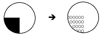
- the above arrangement of receptors can be considered a feature ________ (specifically, for an upper-right-corner)
How is a corner detected?
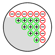
- light falls on the receptors comprising a feature template
- receptors transduce the energy to neural impulses
- activation is sent along, and added together
- if the resulting signal exceeds a (neural) threshold, a corner is “seen”
- thus, the corner has been “made ________” by the visual system
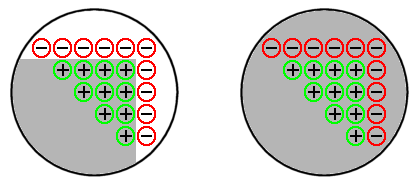
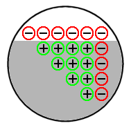
Problem:
- a uniformly bright field, devoid of corners, will also set off receptors, surpassing threshold, and “making explicit” a corner where there is ____!
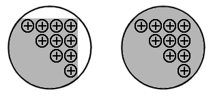
Solution:
- detectors must be more sophisticated
- make some connections __________: when light falls on these receptors, they produce a net negative response:
Problem:
- Is there just ___ upper-right-corner detector in the eye?
- What if the image of an upper-right-corner doesn’t fall on this single detector?
Solution:
- receptive fields on the retina are ___________
- any given receptor is not connected to a single feature detector--but to ____ at once
e.g., any given receptor may be part of arrays trying to detect
• an upper-left-corner
• a horizontal line
• a lower-right-corner, etc.
- in this way, there may be a ______ of feature detectors at each point in space
- one logical implication of such a construction would be a very _______ system (!)
Problem:
- how does our corner-detector handle _____?
- ____--edges cause the detector to exceed threshold!
Solution:
- increase the threshold
Problem:
- if the threshold is increased, ___ corners will not be detected
- is there a satisfactory _____-___?
- nope--template-based systems are intrinsically _________
Conclusion:
- detecting complex features with templates is ______--look for another, better way
As a result of brain damage to a particular region, people may experience a deficit in perception of colour, motion, or faces.
This evidence from _________ reveals that vision is not unitary, but is divided into different structural and functional components.
• Where is this division located?
• What is the nature of the division?
Structure & Function
- early primate visual system has two major pathways:
M pathway: photoreceptors (rods + cones) ![]() diffuse bipolar cells
diffuse bipolar cells ![]() _______ ganglion cells (M-cells)
_______ ganglion cells (M-cells)
P pathway: photoreceptors (mostly cones) ![]() midget bipolar cells
midget bipolar cells ![]() _____ ganglion cells (P-cells)
_____ ganglion cells (P-cells)
- retinal ganglion cell (RGC) anatomical differences:
|
M-cells parasol |
P-cells midget |
|
cell body ____: |
large |
small |
______: |
few (10%) |
many (80%) |
_________: |
sparse, long |
dense, short |
______ from: |
periphery |
fovea |
(____________ type (about 10%): carry signals from short-wavelength cones)
- RGC physiological differences:
|
M-cells parasol |
P-cells midget |
|
conduction _____: |
rapid (15 m/s) |
slow (6 m/s) |
response ____: |
transient |
sustained |
_________ _____ size: |
large |
small |
sensitivity to ________: |
high |
low |
wavelength: |
black & white |
wavelength-sensitive |
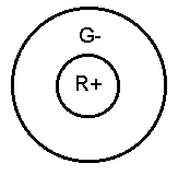
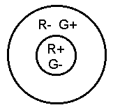
- most ganglion cells project to _______ __________ nucleus (LGN) of the thalamus; has two different major subdivisions:
• _____________: 2 ventral (bottom), large-cell layers
- input comes from large retinal ganglion M-cells
- found in all mammals
• _____________: 4 dorsal (upper), small-cell layers
- input comes from small retinal ganglion P-cells
- found in primates
• _____________: smallest LGN cells located between (and ventral to) parvo and magno layers
- input from bistratified RGCs
- found only in primates
- blue vs. red-green system?
- 10-20% of connections to LGN are from retina; __% come from higher cortical areas; remainder from local inhibitory and brainstem inputs
- LGN is a _______ ______, not simply a relay nucleus
- other major targets for retinal ganglion cell axons include: superior colliculus (initiating visual reflexes), pretectum (pupillary reflexes), accessory optic system (perception of self-motion and gaze stabilization), and suprachiasmatic nucleus (circadian rhythms)
- magno and parvo physiological differences:
1) ______
- magno not differentially sensitive
- parvo (90%) selectively wavelength-sensitive
e.g., colour-opponent centre-surround receptive fields
2) visual ______: ability to detect fine detail
- magno receptive fields are 2× to 3× larger than parvo
- parvo are better at resolving spatial detail
3) _____
- magno respond faster, and have a faster rate of decay than the less-transient parvo cells
- magno better at handling motion
4) ________ sensitivity
- magno are more sensitive to low contrast (1-15% difference in brightness)
- parvo are sensitive to low and high contrast (respond from 1%)
Human Perception
Livingstone & Hubel (1988):
Is there evidence for a ___________ of visual perception?
If so, are these differences what we would expect, given what we know about the magno and parvo systems?
1) ______:
Cavanagh et al. (1984):
- used ____________ stimuli: same intensity, different wavelengths
- motion perception of equiluminant red & green sinewave stripes impaired
Campbell & Maffei (1981):
- slowly rotating disk with stripes of 16 or 32 cycles/ degree often appeared to be __________
- motion perception impaired at high spatial frequencies
2) _____:
Livingstone & Hubel (1987):
- pictorial depth cues rendered with ____________ colours: depth perception eliminated or diminished
- all elements of the image are visible
- implies ___-_____ processes are responsible
3) ______-______ discrimination:
- linking by collinearity (e.g., illusory contours) breaks down with equiluminant stimuli

- may be due to inactivation of depth mechanism
Magno and parvo systems appear to function _______________!
Magno: |
Parvo: |
motion detection |
____ analysis |
________ analysis |
spatial analysis |
depth perception |
______ vision |
more primitive |
recently _______ |
Why this subdivision?
- magno: good for transient object perception
- parvo: better at sustained, detail perception; colour perception can defeat camouflage; is adaptive
- __________ (shape, location, orientation) is adaptive
- ___________ into subsystems allows for greater specialization, and more complex processing of the visual scene
Primary visual cortex, a.k.a. V1, _______ cortex, Brodmann’s (1909) area 17:
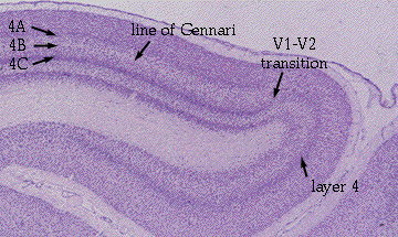
• magno LGN ![]() V1 layer 4Cα
V1 layer 4Cα ![]() 4B
4B ![]() _____ stripes (V2), V3, & MT (medial temporal cortex, a.k.a. V5)
_____ stripes (V2), V3, & MT (medial temporal cortex, a.k.a. V5)
- cells in 4B are ___________ selective, selective for motion in a particular direction, have binocular sensitivity, but no ______ sensitivity
- involved in motion/spatial analysis
• parvo LGN ![]() V1 layer 4Cβ
V1 layer 4Cβ ![]() interblobs (layers 2 & 3)
interblobs (layers 2 & 3) ![]() ____ stripes (V2)
____ stripes (V2) ![]() V4
V4
- involved in processing form
• parvo LGN ![]() V1 layer 4Cβ
V1 layer 4Cβ ![]() blobs (layers 2 & 3)
blobs (layers 2 & 3) ![]() thin stripes (V2)
thin stripes (V2) ![]() V4
V4
- involved in processing colour
• konio LGN ![]() blobs (and other regions including MT)
blobs (and other regions including MT)
• _____: pillar-like cortical sections, found in V1 layers 2 & 3; revealed by cytochrome oxidase staining of mitochondria (Wong-Riley, 1979)
- input from parvo and konio
- no orientation selectivity; have monocular sensitivity; __________ or brightness selective; most are doubly colour-opponent
• __________: areas between blobs in V1 layers 2 & 3
- input from parvo
- cells are ___________ selective; have small receptive fields; have binocular sensitivity
- not wavelength selective, or to direction of movement
David Hubel & Torsten Wiesel (1981 Nobel Prize):
- found cells that responded well to certain stimuli:
• ______ cell (V1: layer 4) responds best to:
- a bar, line, or edge of light
- in a particular location on the retina
- having a specific orientation
- constructed by __________ centre-surround cells?
• _______ cell (V1: layers 2, 3, 5, 6; V2) responds best to:
- a bar, line or edge of light
- in a particular location on the retina
- having a specific orientation
- and moving in a certain _________
- constructed by converging simple cells?
• ____________/end-stopped cell (V2, V3) responds best to:
- a bar, corner, or _____ having a certain length and/or width
- in a particular location on the retina
- having a specific orientation
- moving in a certain direction
- when inserting electrode _____________ to surface of cortex, cells respond to stimuli having the same property
• ________ column: cells respond to stimuli from same retinal location
• ______ _________ column: cells respond to stimuli presented to one eye only
• ___________ column: cells respond to stimuli of the same orientation; adjacent orientation columns differ in orientation selectivity by about 10°
- ___________: 2 mm × 2 mm region containing a single location column, which contains left and right ocular dominance columns, each of which contains the set of orientation columns from 0° to 180°
- the striate cortex has about _____ hypercolumns, which act in parallel
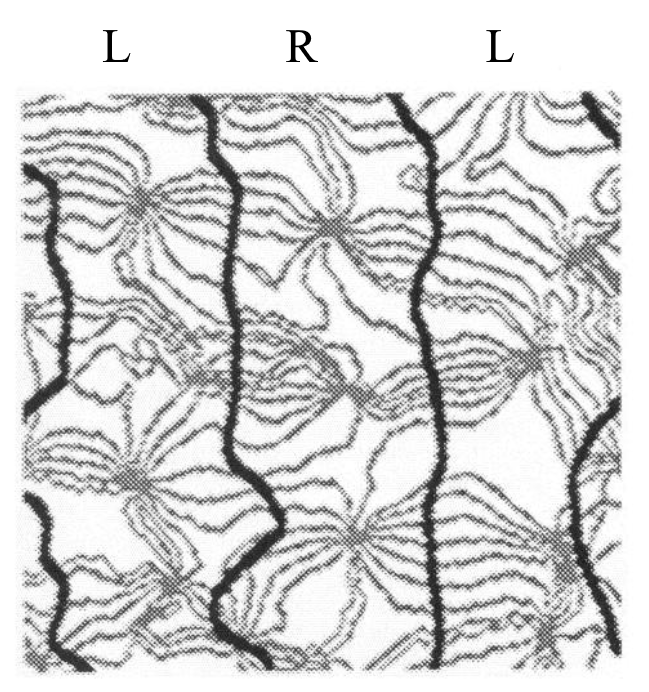
Bonhoeffer & Grinvald (1991):
- hypercolumns are not organized in rectangular blocks or “_____”
- ocular dominance columns form wandering, roughly parallel “_____” (as seen from the surface of the brain), alternating between left and right ocular dominance columns
- developed optical imaging technology with a resolution of 0.05 mm (vs. fMRI’s 1-3 mm):
• applied voltage-sensitive dye which stains cell _________ of striate cortex of monkey
• presented one particular kind of stimulus (e.g., grating of a certain orientation, to one or both eyes)
• ____________ the cortex
• repeated with different stimuli
• superimposed the images
- pictures colour-enhanced by computer: each orientation column given a colour; similar ____________ have similar colours
- particular (peculiar?) patterns of orientation columns were revealed within a given ocular dominance column
- surface of the columns forms “_________”
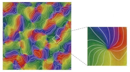
Obermayer & Blasdel (1993):
- described pinwheel structure
- iso-orientation contours (or “______”, lines that represent cells having the same orientation preference) radiate from “_____________” lying in the centre of the ocular dominance bands
- at a singularity, all the different orientations meet; singularities are centred above blobs? (no, just a coincidence)
- spokes of the pinwheel cross the borders between left and right ocular dominance bands at right angles
- some pinwheels have spokes that radiate iso-orientation contours clockwise (-); some are counter-clockwise (+); spokes differ by about 10° orientation
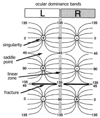
- ______ ____ between bands comprised of binocular cells
- “______ ______” exist between two singularities of the same sign; orientation changes slowly in this region
- “_________” exist between singularities of opposite signs; indicate a sharp change in orientations
- structural organization of orientation-selective neurons in the cortex is ______
- the retina does not map to the cortex in an isomorphic point-to-point fashion
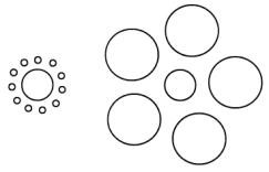
The Packing Problem
- the job of the visual cortex is to take information that is implicit in the incoming information, and make it explicit
- all of this newly explicit information must be coded in neurons
- the visual cortex encodes data (e.g., lines) that have three parameters of information: X position, Y position, and orientation
- although the cortex is three dimensional, cells within a column all respond to the same orientation--so cortical depth is not used to encode different orientations
- the packing problem: how are three parameters of information mapped onto a 2-D representational space?
- solving this problem of mismatching dimensions mathematically produces _____________
e.g., fingerprints have singularities because they are mapping one parameter (orientation) onto a 2-D space (the skin); this is a mismatch the opposite way (1-D ![]() 2-D)
2-D)
- cortical depth does encode other stimulus aspects at different cortical levels, like motion (simple vs. complex cells), etc.
Two extrastriate streams in vision:
• dorsal stream/parietal pathway/“_____” or “how”
- receives mostly _____ input
- neurons in area MT/V5 appear to be involved in analyzing speed and direction of stimulus motion, and stereoscopic depth analysis; implicated in ___________: inability to perceive motion of objects
- neurons in area MST may be involved in visual navigation, directing eye movements, and motion perception
- projects to posterior ________ association cortex
- this stream seems to be processing aspects of the ________ of visual stimuli
• ventral stream/inferotemporal pathway/“____”
- receives mix of magno and parvo
- V4 involved in colour processing; implicated in cerebral _____________: deficit in colour vision without loss of object perception
- V4 cells also orientation selective, suggesting form analysis
- IT (inferior temporal) involved in processing complex spatial arrangements; implicated in _____________: loss of ability to recognize faces with otherwise normal vision
- projects to V2-V5, inferior temporal cortex (TEO, TE, superior temporal sulcus [STS])
- this stream seems to be processing _____ and ______
Evidence for this segregation:
• single-cell recording and brain imaging
• Balint’s syndrome (Rudolf Bálint, 1909):
- patient had bilateral damage to superior posterior parietal lobes (very rare)
- optic ataxia: inability to reach for and _____ objects in field of view
- optic apraxia: inability to guide eye movements or change visual fixation
- _______________: inability to perceive more than one aspect of a visual stimulus and integrate details into coherent whole
- could recognize individual objects, but could not tell where they were located or reach for them
- intact “what” system, but damaged “where” system?
• Visual form _______:
- inability to extract global structure, despite intact low-level sensory processing (acuity, colour, and brightness discrimination intact)
- cannot recognize, discriminate, or copy complex visual forms, like shapes
- but they can _____ objects they cannot identify
- intact “where” system, but damaged “what” system?
• Aglioti, DeSouza, & Goodale (1995):
- built tactile version of Ebbinghaus illusion (a.k.a. Tichener circles) out of things like poker chips
- showed observers the illusion:
• visually, the ____ of the centre disk was affected by the surrounding disks (even though both centre disks were equal in size)
- observers were then asked to pick up the centre disk
• cameras tracked infrared LEDs to measure grip aperture (opening between index finger and thumb)
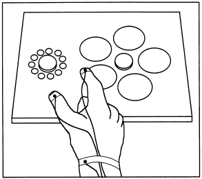
• grip aperture was perfectly calibrated to the exact size of the disk--well before contact with the disk
• that is, observers treated disks that were perceptually different as physically _________
- conclusion: visual perception and visually guided action are ________
This evidence shows a double-dissociation of extrastriate pathways:
• how to determine whether two related functions are independent?
• in one person with brain damage, function 1 is impaired, but function 2 is intact
• and in another person with brain damage, function 1 is intact, but function 2 is impaired
• Balint’s syndrome and visual form agnosia show a double dissociation of what vs. where
• Aglioti et al. (1995) showed a double dissociation within intact brains
Why do we have two complementary extrastriate streams?
• dorsal stream: specialized to ________ with objects (same every time)
e.g., picking up a mug of beer is the same every time
• ventral stream: specialized to ________ objects from various POVs (shape constancy)
e.g., seeing a mug of beer from different angles, different retinal images